Structural Classification of Membrane Proteins
Membrane proteins represent about a third of the proteins in living organisms. Based on their structure, there are main three types of membrane proteins: the first one is integral membrane protein that is permanently anchored or part of the membrane, the second type is peripheral membrane protein that is only temporarily attached to the lipid bilayer or to other integral proteins, and the third one is lipid-anchored proteins (Fig. 1). Here we only describe the first two types of membrane protein.

Fig. 1 Structural classification of membrane proteins
According to their their relationship with the bilayer, integral membrane protein can be classified two primary types: integral polytopic proteins and Integral monotopic proteins. Integral polytopic proteins are also known as “transmembrane proteins” which can span across the membrane at least once (Fig. 2). These integral membrane proteins may have different transmembrane topology which refers to orientations (locations of N- and C-termini) of membrane-spanning segments with respect to the inner or outer sides of the biological membrane occupied by the protein. The first three types in the Fig. 2 are common forms in integral membrane proteins, such as, transmembrane α-helix protein, transmembrane α-helical protein and transmembrane β-sheet protein.
Integral monotopic proteins are one type of integral membrane proteins that are attached to only one side of the membrane and do not span the whole way across. There are 4 types of interaction between Integral monotopic membrane protein and cell membranes: by an amphipathicα-helix paralle, by a hydrophobic loop, by a covalently bound membrane lipid and electrostatic or ionic interaction with membrane lipids (No. 4, 5, 6,7 of Fig. 2). Integral membrane proteins can be separated from the biological membranes only using detergents, nonpolar solvents, or sometimes denaturing agents. Peripheral proteins dissociate following treatment with a polar reagent, such as a solution with an elevated pH or high salt concentrations.

Fig. 2 Seven types of Integral Membrane protein Structure
The membrane is represented in yellow. 1. A single transmembrane α-helix (bitopic membrane protein). 2. A polytopic transmembrane α-helical protein. 3. A polytopic transmembrane β-sheet protein. 4. Interaction by an amphipathic α-helix parallel to the membrane plane (in-plane membrane helix). 5. Interaction by a hydrophobic loop. 6. Interaction by a covalently bound membrane lipid. 7. Ionic or electrostatic interactions with membrane lipids.
Function of Membrane Proteins
Membrane proteins play crucial roles in all organisms, where they serve as, such as membrane receptors, ion channels, GPCR (G protein–coupled receptors) and various kinds of transport proteins. Membrane receptors that embedded in the cell membranes, which can transmit signals between the cell’s internal and external environments. Membrane transport protein (or transporter) is a kind of membrane protein that involved in the movement of ions, small molecules, or macromolecules across a biological membrane. About membrane transport protein, there is a detailed classification called Transporter Classification Database (or TCDB) approved by International Union of Biochemistry and Molecular Biology. Ion channels are one of most important type of membrane transport proteins. The functions of ion channel include establishing a resting membrane potential, shaping action potentials and other electrical signals by gating the flow of ions across the cell membrane, controlling the flow of ions across secretory and epithelial cells, and regulating cell volume. Some enzymes are also membranes proteins, for example oxidoreductase, transferase or hydrolase. Cell adhesion molecules that located on the cell surface involved in binding with other cells or with the extracellular matrix (ECM), allow cells to identify each other and interact. For example, proteins including Ig (immunoglobulin) superfamily involved in immune response. The following figure 3 summarizes membrane protein functions for easy to understand.

Fig. 3 Membrane proteins functions
Expression of Membrane Proteins
Owing to their central role in basically all physiological processes, membrane proteins constitute around 60% of approved drug targets, and, therefore, their experimentally determined three – dimensional structures are eagerly sought to assist in structure – based drug design. Despite significant and considerable recent improvements, the expression of functionally folded membrane proteins in sufficient amounts for functional and structural studies is still a challenging task. Compared with soluble cytoplasmic proteins, a variety of difficulties in expression of membrane proteins. 1. Membrane proteins are not just released into the cytosol but must rather be targeted and translocated to their final destinations in membranes. In particular in eukaryotic cells, there is need a more complicated biological process requires sophisticated recognition and sorting mechanisms. 2. Copy number and capacity of prokaryotic and eukaryotic expression systems are limited because of translocation machineries, and translocation can be selective only for distinct groups of membrane proteins. 3. For structural and functional studies or other purposes, it is have to extract membrane protein from cellular after expression, followed by transfer into artificial and defined hydrophobic environments like micelles or liposomes. In this procedure, it is highly critical as membranes have to be disintegrated by relatively harsh detergents which can result in conformational aberrations or in unfolding of membrane proteins. It is therefore not surprising that the structural information obtained from membrane proteins with some 150 unique structures and only ~7% entries in the Protein Data Bank is lacking far behind the data obtained from soluble proteins.
Core factors that determine the yield, integrity, activity and stability of synthesized membrane proteins mainly are the availability of highly processive transcription and translation machineries, suitable folding environments, the lipid composition of cellular membranes, the presence of efficient targeting systems and appropriate pathways for posttranslational modifications (PTMs). Expression of membrane proteins in expression systems/cellular background as closely related to their origin as possible may be the most reasonable strategy. However, the efficiency and workload of the individual expression systems are quite different, and preparative amounts of membrane proteins are often only obtained with bacterial, yeast or cell-free systems (Fig. 4).
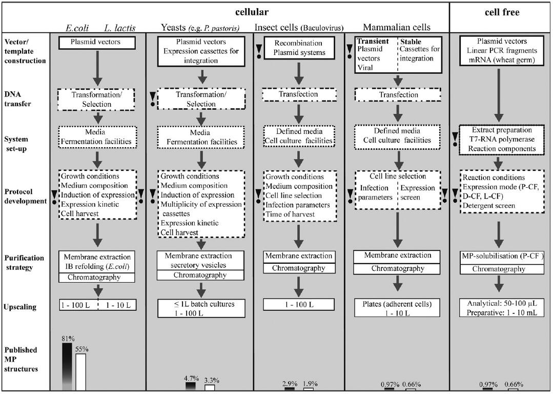
Fig. 4 Process design of membrane protein expression systems.
Overview of protocol development for the most popular membrane protein expression systems. Basic optimization parameters characteristic for the individual expression systems are illustrated, and the most critical parts are indicated. Some optimization parameters are not considered: vector or target design, e.g. fusion technologies or modifications of the expression cassettes. Grey bars illustrate the success in obtaining membrane protein structures after using the individual expression systems, while white bars correspond to those membrane protein structures obtained only after recombinant expression. (F. Junge et al.)
Although choosing an appropriate expression system need to consider many factors which may increase the complexity and time-consuming of research project, Creative Biolabs can provide comprehensive membrane protein production service which can help you decide optimization parameter and systems for your targeted experiments.
Membrane Protein Antibody
Membrane proteins represent the vast majority of clinical drug targets and are a focus of pharmaceutical and biotechnological interests. Despite significant and considerable recent improvements, the expression of functionally folded membrane proteins in sufficient amounts for functional and structural studies is still a challenging task. However, there are still many strategies for preparing membrane proteins that have been developed by Specialists from Creative Biolabs. In terms of anti-membrane protein antibody discovery and production, Creative Biolabs is able to provide various methods from immune antibody library construction by phage display to native antibody discovery by antigen-specific B lymphocytes cytometry technology. If you are interested in discovering novel membrane protein antibodies, please feel free to contact us for more details.