Cell proliferation is an important vital sign of the organism in a splitting manner. Single-celled organisms produce new individuals in the form of cell division, while a multicellular organism that produces new cells in a cell division to replenish aging and dying cells. Cellular organisms can be developed by a fertilized egg, through cell division and differentiation, and eventually, develop into a new multicellular individual. The transferred genetic material can be distributed equally among the two daughter cells by cell division.
It can be proved that cell proliferation is the basis for the growth, development, reproduction, and inheritance of organisms, while the purpose of T cell proliferation is to provide dynamic monitoring of specific immunotherapy in patients with HBV for liver disease clinicians and to evaluate efficacy at the end of treatment.
The principle of T cell proliferation is that peripheral blood mononuclear cells (PBMC) in patients with chronic HBV infection are incubated with HBV epitope polypeptides and cytokines for 1 week. T cells in patient peripheral blood will proliferate under the stimulation of HBV epitope peptide antigens. The number of active cells was detected by MTT method to calculate the value of SI.
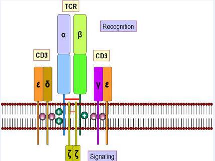 The T-cell receptor, or TCR, is a molecule found on the surface of T cells. A few TCR molecules of mature T cells is a heterodimeric molecule consisting of a gamma chain and a delta chain, similar in structure to TCRαβ, also known as TCR-1. It can directly identify antigens without combining with MHC and antigen presenting molecules. After the recognition of pathogen surface antigen molecules, based on T cell culture, TCR will proliferate and differentiate into the effector cell to play a role in the damaging effect, while the cells and tumor cells infected with the virus will have a cytotoxic effect.
T cell proliferation assay, also known as T cell transformation test, can be accomplished through cell changes and cytoplasmic expansion on the type of morphological method and nuclide method. T cells are stimulated in vitro by a certain material, then cell metabolism and morphological are changed continually to increase the protein and nucleic acid synthesis in 24~48h, resulting in a series of changes in proliferation, such as cell changes, cytoplasm expansion, vacuoles, nuclei Ren obvious, nuclear chromatin loose, etc. Lymphocytes will change into lymphoblasts. Therefore, this lymphocyte proliferation is also called lymphocyte transformation, which can determine the reactivity and functional status of the lymphocytes.
The non-antigenic stimuli include such as phytohemagglutinin (PHA), concanavalin A (ConA), American land (PWM), lipopolysaccharide (LPS), commonly known as the mitogen. LPS stimulates B cells; PWM stimulates T and B cells; PHA and ConA stimulate T cell proliferation.
On the other hand, antigenic stimuli contain such as tuberculin, staphylococcal toxin, tetanus toxoid, streptococcal kinase, tumor antigen, allogeneic tissue antigen etc.
There are two experimental types of T cell proliferation, morphological method, and radionuclide method. The principle of the morphological method is to mix peripheral blood or separated single cells with certain phytohemagglutinin (PHA) with a culture of 72h under 37 ℃, taking the culture cells for smear microscopy. Depending on cell sizes, the ratio of nucleus to the cytoplasm, the staining and nuclear structure of the cytoplasm, and the presence or absence of nucleolus, lymphocytes, transitional mother cells and mature small lymphocytes in mitochondrial phases will be counted respectively for 200 cells to calculate the conversion rate.
As for the radionuclide method, the vast majority of peripheral blood T cells in the cell cycle of the G0 period are activated by specific antigens or mitogens, stepping from the G0 period into the G1 phase. Then proteins, RNA and DNA precursors substances are synthesized to prepare the material basis for DNA replication. Next, in the S phase, the amount of cell synthesis DNA will double and the cell proliferation degree can be speculated by the amount of 3H labeled TDR.