Immune Checkpoint Inhibitor-expressing Oncolytic Virus
With years of exploration in oncolytic virus development, Creative Biolabs has built an advanced oncolytic virus engineering platform. With our excellent scientists, we can provide high quality immune checkpoint inhibitor-expressing oncolytic virus to meet customers' requirements. We are dedicated to designing and developing desirable oncolytic virus to contribute greatly to your research, preclinical study and drug development.
Oncolytic viruses are a group of replication competent viruses which curtail tumor growth. They are able to selectively replicate within tumor cells and lead to cell death while leaving normal cells intact, on account of their natural capacity or via genetic modifications. Oncolytic viruses also can cause tumor cell death through the induction of innate and adaptive immune responses. Nowadays, the identification and targeting of immune checkpoints is becoming a novel approach that has the capacity to activate sustained antitumor immunity. Antibodies blocking checkpoint proteins have been approved to treat cancer, including anti-programmed cell death-1 (PD-1) and anti-cytotoxic T-lymphocyte antigen-4 (CTLA-4). Creative Biolabs offers high quality immune checkpoint inhibitor-expressing oncolytic virus to contribute greatly to your research, preclinical study and drug development.
CTLA-4 and PD-1 Checkpoint Molecules
CTLA-4 and PD-1 are two extensively studied inhibitory immune molecules, sharing some similar characteristics. However, their mechanism of the action to limit T cell activation are different. As T cells are activated, CTLA-4 is a major intracellular protein that translocates to the immunological synapse and co-localizes with TCRs and lipid rafts. CTLA-4 is able to restrict conjugation times between T cells and APCs and enables to change the signaling pathway to limit T cell proliferation and cytokine generation. It serves an essential part in maintaining immune regulated homeostasis via exerting its effects mainly through down regulating CD4 T helper T cells functions, as well as promoting suppressive functions of Tregs. PD-1 is a transmembrane protein functioned on resting naïve and memory T cells. It has two ligands and is expressed on activated T cells upon TCR engagement. PD-1 regulates T cell suppression via binding to its ligands, PD-L1 and PD-L2, to transmit inhibitory signals to impair T cell effector functions.
 Fig 1. Schematic diagram of the programmed cell death 1 (PD-1)/PD-L1 pathway. (Santarpia, M., 2015)
Oncolytic Viruses with Anti-CTLA-4 Therapy
In the past studies, researchers have provided pre-clinical data to support clinical study of the use of checkpoint antibodies with oncolytic Newcastle disease virus (NDV). They employed a clinically relevant pre-clinical model of metastatic tumor and showed that the combination therapy of CTLA-4 checkpoint blockaded and NDV controlled both local and distant tumors better than either NDV or anti-CTLA-4 treatment alone. The combination approach also resulted in long-term survival of mice, elicited inflammatory recruitment of CD8 T cells, and induced overall improvement of effector to Tregs ratio. Moreover, it has demonstrated that the combination therapy provides better protection against tumor challenge and contributes to a robust memory response.
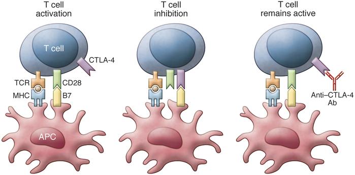 Fig 2. T cell activation needs costimulation via TCR and CD28. Binding of B7 to CTLA-4 inhibits T cell function. Anti–CTLA-4 antibodies block CTLA-4 binding and prevent inhibition of T cell function. (Buchbinder, E., 2015)
Oncolytic Viruses with Anti-PD-1 Therapy
Reovirus, a double stranded RNA virus, has direct oncolytic activity against a number of tumor types. Rajani et al. have exhibited that combination of intra-tumoral reovirus (Reolysin) with systemic anti-PD-1 antibody delivery was far superior in establishing tumor control than either of the monotherapies. Seven days after the first reovirus administration, anti-PD-1 antibody is treated to minimize induction of anti-viral response and allows the virus to spread within the tumor. Moreover, depleting CD8 T cells and NK cells completed ablated the efficacy of therapy, in favor of the interplay of both innate and adaptive immune system to mediate clinical efficacy.
With our well-established oncolytic virus engineering platform, the experienced scientists at Creative Biolabs offer high quality oncolytic virus expressing immune checkpoint inhibitor, such as B & T lymphocyte attenuator and antibodies targeting checkpoint molecules, including CTLA-4, PD-1, TIM-3, LAG-3. We also provide other various services regarding oncolytic virus development. Please feel free to contact us for more information and a detailed quote.
Other Services in Oncolytic Virus Engineering Platform
- Pathogenicity Manipulation (Attenuation)
- Immunogenicity Manipulation
- Antibody-expressing Oncolytic Virus
- Cytokine/Chemokine-expressing Oncolytic Virus
Reference:
- Buchbinder, E., (2015). "Cytotoxic T lymphocyte antigen-4 and immune checkpoint blockade." The Journal of clinical investigation, 125(9), 3377.
- Santarpia, M., (2015). "Tumor immune microenvironment characterization and response to anti-PD-1 therapy." Cancer biology & medicine, 12(2), 74.
- Rajani, K. R.,. (2015). "Harnessing the power of onco-immunotherapy with checkpoint inhibitors." Viruses, 7(11), 5889-5901.