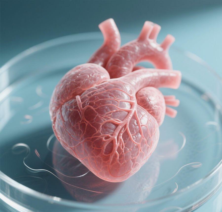
Cardiac Organoids Introduction
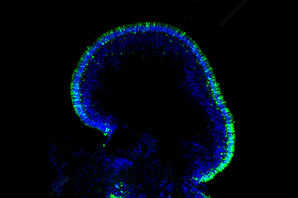
Cardiac organoids are miniature, self-organizing 3D structures derived from pluripotent stem cells (PSCs) or adult stem cells that closely mimic the cellular composition, structural organization, and functional characteristics of native heart tissue. Cardiac organoids evolve into intricate microtissues with multiple cardiac cell types like cardiomyocytes, endothelial cells, and cardiac fibroblasts which organize themselves into spatial patterns that resemble physiological conditions unlike 2D cultures. The self-assembly mechanism of cardiac organoids replicates fundamental aspects of embryonic heart formation which provides a novel perspective on human heart development and disease. Developing "mini-hearts in a dish" significantly affects basic research while also advancing drug discovery and regenerative medicine fields.
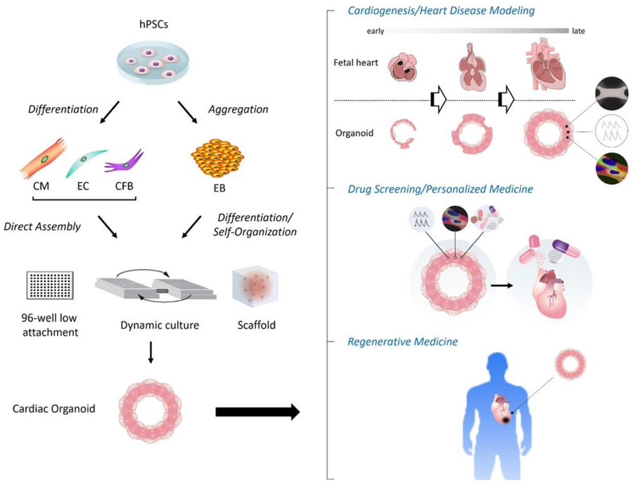 Figure 1 Applications of human cardiac organoids.1,3
Cardiac organoids-on-a-chip represent a cutting-edge fusion of three-dimensional (3D) cardiac organoid technology and microfluidic "organ-on-a-chip" (OoC) systems. The innovative platforms simulate human heart complexities in structure, function, and physiology to provide unparalleled insights into cardiac development research along with disease modeling and drug screening applications in regenerative medicine.
Cardiac organoids-on-a-chip integrate two revolutionary technologies:
Cardiac organoids
Clusters form when pluripotent stem cells and cardiac progenitor cells self-organize into three-dimensional cell structures. These formations integrate cardiomyocytes with endothelial cells and fibroblasts to replicate early cardiac tissue and they show the ability to contract spontaneously and produce electrical signals.
Organ-on-a-chip (OoC)
Microengineered devices contain living cells which cover micron-scale channels while perfused culture media replicates blood flow and mechanical forces including cyclic stretch. Organ-on-a-chip systems replicate tissue-tissue interfaces like blood-tissue barriers along with physiological dynamics which static 2D cultures cannot replicate.
Cardiac Organoid Culture
Creating cardiac organoids requires an extremely precise environment that mimics the heart's natural surroundings. The cultivation process uses specialized culture media alongside specific matrices and growth factors. Cardiomyocyte differentiation and function modulation necessitates specialized culture media which contains essential nutrients, vitamins, minerals, growth factors along with optional small molecules or drugs. Collagen and synthetic hydrogels along with Matrigel serve as physical frameworks that support cell adherence and expansion. Essential growth factors such as cytokines and heart-specific hormones stimulate PSCs and CPCs to develop into cardiomyocytes while maintaining cardiac organoid health and viability during culture.
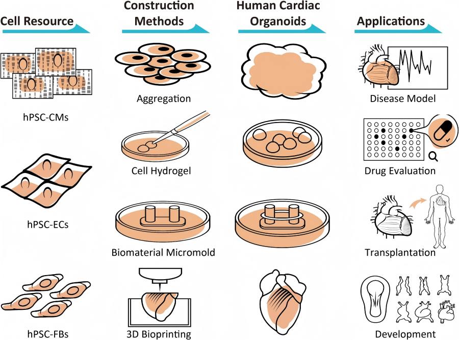 Figure 2 Construction methods of human cardiac organoids.2,3
Cardiac Organoid Cell Sources: iPSCs vs. ESCs
|
Parameter
|
iPSCs
|
ESCs
|
|
Origin
|
Patient somatic cells (autologous)
|
Embryonic blastocysts
|
|
Ethics
|
No ethical concerns
|
Controversial
|
|
Differential Efficiency
|
Higher for disease modeling (e.g., hypertrophic cardiomyopathy)
|
Standard for developmental studies
|
|
Limitations
|
Epigenetic memory; teratoma risk
|
Immune rejection
|
Electrophysiological Property Evaluation of Cardiac Organoids
The use of Microelectrode array (MEA) technology forms the fundamental basis for evaluating cardiac organoids' electrophysiological characteristics. The MEA system allows simultaneous detection of multiple electrophysiological signals in real-time thanks to its multiple electrodes integrated into a single chip. The method demonstrates both high-throughput capacity and precise results while remaining adaptable for different biological samples such as cells, tissue slices and organoids. MEA demonstrates superior performance in cardiac research by accurately recording essential functional activities including electrical signal conduction as well as field potentials, contractions and action potentials. The capability provides essential support for fast compound screening procedures and toxicology surveillance while helping to study alterations in conduction velocity and arrhythmic events.
Applications of Cardiac Organoids
Cardiac organoids have emerged as powerful tools with diverse applications across basic and translational research:
Disease Modeling
-
Congenital Heart Defects: Modeling congenital heart defects requires recapitulating early developmental processes to understand their underlying mechanisms.
-
Cardiomyopathies: Cardiac organoids generated from patient-specific iPSCs serve as models for genetic cardiomyopathies and help discover new therapeutic targets.
-
Arrhythmias: The electrophysiological characteristics of organoids serve as valuable tools for investigating arrhythmia mechanisms and evaluating anti-arrhythmic medications.
-
Ischemic Heart Disease: The study of cell death and regeneration after hypoxic and reperfusion injuries relies on modeling these processes in a 3D context.
-
Cardiotoxicity Assessment: Early-stage drug development now includes high-throughput screening for cardiotoxic effects which helps to reduce expensive clinical trial failures by identifying harmful drug candidates sooner.
-
Drug Efficacy Testing: The therapeutic potential of new compounds is assessed by examining their effects on cardiac function and disease phenotypes.
Developmental Biology
-
Studying Cardiogenesis: Unraveling the complex signaling pathways and cellular interactions that govern heart development.
-
Teratogen Screening: The investigation aims to pinpoint environmental substances or pharmaceutical agents that may lead to heart developmental disorders in prenatal stages.
-
Cell Therapy Research: Serving as a platform to test strategies for enhancing the engraftment, survival, and integration of transplanted cardiac cells.
-
Bioengineering Heart Tissue: The technology supplies fundamental components for constructing larger engineered cardiac tissues that can be used for transplantation and sophisticated in vitro models.
Frequently Asked Questions of Cardiac Organoids
Q: What are cardiac organoids and how do they differ from 2D cell cultures?
A: Cardiac organoids represent small 3D tissue constructs grown from stem cells in laboratory conditions that replicate both the cellular structure and cardiac function of the human heart. In contrast to 2D cell cultures that develop on flat surfaces organoids enable cells to form interactions within a physiological three-dimensional space which promotes complex tissue organization and functionality including spontaneous beating and electrophysiological activity through cell-cell and cell-ECM interactions.
Q: What types of stem cells are used to generate cardiac organoids?
A: Researchers commonly employ human pluripotent stem cells (hPSCs), especially human induced pluripotent stem cells (hiPSCs) and human embryonic stem cells (hESCs), for their experiments. While cardiac progenitor cells and mesenchymal stem cells from adults can serve this purpose their differentiation protocols are distinct.
Q: How long does it take to generate functional cardiac organoids?
A: Generating cardiac organoids requires differing amounts of time based on the chosen protocol and the intended level of maturation. The process of initially differentiating cells into organoids that exhibit spontaneous beating takes about 8-10 days. Organoids require several weeks to months of culture time to develop their complex features beyond initial maturation.
Q: Is it possible to generate patient-specific cardiac organoids?
A: Patient-specific cardiac organoids can be produced using induced pluripotent stem cells which originate from the patient's own somatic cells such as skin fibroblasts or blood cells. The models accurately reproduce both the patient's genetic background and disease phenotype which makes them essential for precision medicine and targeted drug testing.
Conclusion
Cardiac organoids mark a revolutionary breakthrough in biomedical research. The 3D mini-hearts created from human pluripotent stem cells emulate major cardiac functions and pathologies which makes them valuable for drug screening applications and regenerative medicine studies. Despite existing obstacles in heterogeneity, scalability and maturation cardiac organoids demonstrate great potential. Advancements in culture protocols along with bioengineering and personalized medicine approaches make cardiac organoids a revolutionary tool for heart disease understanding and treatment.
Transform Your Research Using Cardiac Organoid Models from Creative Biolabs
Creative Biolabs strives to expand the limits of 3D biology by delivering innovative and dependable solutions to overcome major obstacles faced in cardiovascular research and drug development. Our partnership will help you fully harness cardiac organoids' potential to achieve your next research breakthrough.
Organoid Models
Organoids Related Products
Creative Biolabs's comprehensive suite of services, from custom organoid generation and characterization to advanced disease modeling and drug screening, empowers researchers and pharmaceutical companies to accelerate their biomedical discoveries and translate them into tangible clinical benefits. Contact us today to learn more!
Research Model
Related Sections: