Creative Biolabs is offering the most comprehensive services for antibody development projects. With strict regulation and effective execution, we are dedicated to providing the most valuable solutions to complete your projects.
HighlightsCreative Biolabs is offering the most comprehensive services for antibody development projects. With strict regulation and effective execution, we are dedicated to providing the most valuable solutions to complete your projects.
HighlightsCreative Biolabs has established a broad range of platforms for developing novel antibodies or equivalents. These cutting-edge technologies enable our scientists to meet your demands from different aspects and tailor the most appropriate solution that contributes to the success of your projects.
HighlightsWith deep understanding in antibody-related realms and extensive project experience, Creative Biolabs offers a variety of references to help you learn more about our capacities and achievements, including infographic, flyer, case study, peer-reviewed publications, and all kinds of knowledge that can assist your projects. You are also welcome to contact us directly for more specific solutions.
HighlightsGet a real taste of Creative Biolabs, one of the most professional custom service providers in the world. We are committed to providing highly customized comprehensive solutions with the best quality to advance your projects.
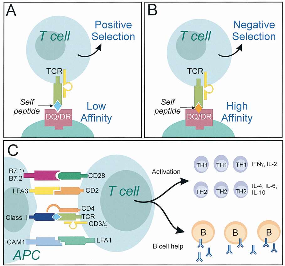
HighlightsCreative Biolabs provides sensitive, specific and accurate SIAT® Naïve T cell proliferation assays to assess immunogenicity, by identifying the epitope sequences that can provoke the proliferation of helper T cells, so that to induce a helper T cell immune response.
Our SIAT® Naïve T cell proliferation assay is an unique, sensitive and accurate approach for immunogenicity assessment, as it utilizes powerful flow cytometry methods, which can accurately determine the percentage of proliferating CD4+ cells and phenotype the T cell responses in details, all with significantly improved overall sensitivity.
Our SIAT® Naïve T cell proliferation assay has been developed to identify the presence or absence of potential T cell epitopes. Thus, it can be applied as a useful tool in preclinical discovery of novel peptides or proteins. It is so sensitive, that can even address the relative immunogenicity between structurally similar molecules. It has been successfully applied to help our clients to distinguish between different candidates, or generate indications for succeeding protein engineering strategies in deimmunization of a potential immunotherapeutic.
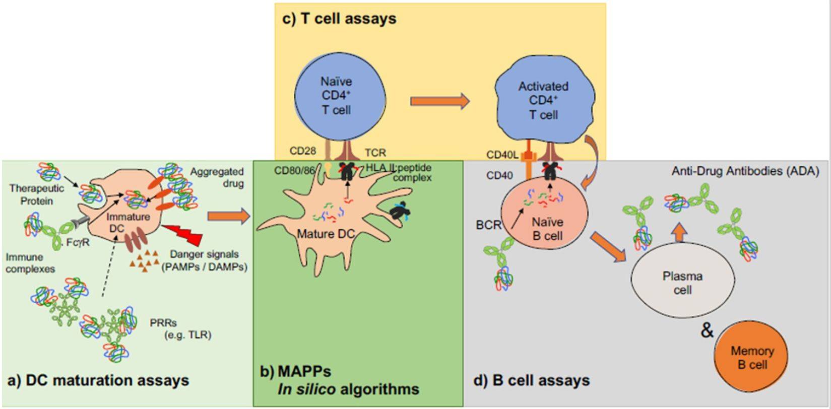
As part of Creative Biolabs SIAT® Immunogenicity System, the T cell proliferation assay can be used for analysis alongside the outcome from cell-free HLA-peptide binding assays for more than 50 class II HLA alleles, for a detailed profile of the helper T cell immune response to one or more drug candidates.
A naïve T cell proliferation assay is a laboratory technique used to measure the activation and division of naïve T cells in response to an antigen, including biotherapeutic drugs. By tracking proliferation, this assay assesses the potential immunogenicity of a substance by simulating an immune response in a controlled environment.
Naïve T cells are used because they have not yet encountered their specific antigen and thus provide a clean slate for assessing the immunogenic potential of new drugs. Their response can indicate how the immune system might react upon first exposure to a biotherapeutic agent.
Flow cytometry allows for the precise measurement of cell proliferation and other parameters like cell size, granularity, and protein expression by using fluorescently labeled antibodies that bind to specific cell markers. This enables detailed analysis of T cell activation and proliferation in response to antigens.
Common markers include Ki-67, a protein expressed during cell proliferation, and CFSE (carboxyfluorescein succinimidyl ester), a dye that dilutes as cells divide. Flow cytometry tracks these markers to quantitatively measure how many cells are dividing in response to the drug tested.
The assay setup involves isolating naïve T cells, labeling them with proliferation markers, exposing them to the drug or antigen, and then culturing them under conditions that support cell growth. After a designated period, the cells are analyzed by flow cytometry to assess proliferation.
While the assay provides valuable insights into how a biotherapeutic might be perceived by the immune system, it does not fully replicate the complexity of human immunity. Results can guide further development and testing but should be corroborated with clinical data for accurate immunogenicity prediction.
Limitations include variability in T cell sources, the artificial nature of in vitro conditions, and potential discrepancies between in vitro and in vivo responses. Additionally, some biotherapeutics may require co-stimulatory signals or accessory cells to elicit a full immune response, which can be challenging to replicate precisely in vitro.
The duration of the assay varies but typically spans from several days to a week. This period allows adequate time for cell activation, proliferation, and subsequent analysis by flow cytometry.
Use the resources in our library to help you understand your options and make critical decisions for your study.
All listed services and products are For Research Use Only. Do Not use in any diagnostic or therapeutic applications.