Anti-Fucosyltransferase (FUT) Antibody Development Service
Creative Biolabs stands at the forefront of glyco-immunology, offering specialized solutions for the detection and analysis of enzymes responsible for glycan synthesis. As a key part of our Anti-Glycan Related Enzyme Antibody Development Service, we present a comprehensive platform for developing high-specificity antibodies against the entire human fucosyltransferase (FUT) family. This service encompasses all 13 identified human FUTs (FUT1-FUT9 and POFUT1-POFUT4), supporting pivotal research in cancer biology, immunology, and developmental signaling. By generating isoform-specific antibodies that distinguish between closely related enzymes (e.g., FUT1 vs. FUT2, or FUT3-7), we empower researchers to dissect the complex roles of aberrant fucosylation in malignancy and inflammation, paving the way for the development of next-generation serological detection tools and therapeutic strategies.
Introduction to Fucosyltransferases: A Diverse Enzyme Family
Fucosylation is a biologically significant glycosylation event involving the transfer of an L-fucose residue from GDP-fucose to oligosaccharides, glycoproteins, or glycolipids. This process is catalyzed by fucosyltransferases (FUTs), which are categorized based on the linkage they form (alpha-1,2, alpha-1,3/4, alpha-1,6, or O-linked). In humans, these enzymes are critical for processes ranging from ABO blood group determination to leukocyte trafficking and Notch signaling. The structural diversity and specific expression patterns of these enzymes make them high-value targets for research into disease mechanisms.
Fucosyltransferases are generally classified into four subfamilies. The alpha-1,2-FUTs (FUT1, FUT2) determine the H-antigen status; the alpha-1,3/4-FUTs (FUT3-7, FUT9) synthesize Lewis antigens; the alpha-1,6-FUT (FUT8) is responsible for core fucosylation of N-glycans; and the Protein O-fucosyltransferases (POFUT1-4) modify specific protein domains in the endoplasmic reticulum (ER). Understanding the specific substrates and biological roles of each is essential for selecting the right antibody target.
| Subfamily | Enzymes | Linkage / Specificity | Primary Substrates & Products | Key Biological Roles |
|---|---|---|---|---|
| alpha-1,2-FUTs | FUT1 | Gal-β1,4-GlcNAc (α1,2) | Type 2 Lactosamine → H-antigen | Erythrocyte/Endothelial adhesion; Angiogenesis |
| FUT2 | Gal-β1,3-GlcNAc (α1,2) | Type 1 Lactosamine → H-antigen (Secretor) | Mucosal defense; Gut microbiota regulation | |
| alpha-1,3/4-FUTs | FUT3 | α1,3 and α1,4 | LeA, LeB, sLeA, sLeX | Cancer metastasis (GI tract); E-selectin ligand synthesis |
| FUT4 | α1,3 | LeX, sLeX (myeloid cells) | Embryonic adhesion; Leukocyte recruitment | |
| FUT5 | α1,3 and α1,4 | sLeA, sLeX | Fertilization; Cancer cell migration | |
| FUT6 | α1,3 | sLeX (plasma/liver) | Plasma FUT activity; Liver metastasis | |
| FUT7 | α1,3 (Sialylated only) | sLeX (Leukocytes) | Principal regulator of leukocyte extravasation (inflammation) | |
| FUT9 | α1,3 | LeX (Neural/Brain) | Neural development; Neurite outgrowth | |
| alpha-1,6-FUT | FUT8 | Core GlcNAc (α1,6) | N-glycan Core Fucose | Receptor signaling (EGFR, TGF-β); IgG ADCC modulation |
|
POFUTs (ER Resident) |
POFUT1 | O-Fucose on Ser/Thr | EGF-like domains (e.g., Notch) | Notch signaling activation; Developmental fate |
| POFUT2 | O-Fucose on Ser/Thr | Thrombospondin Type 1 Repeats (TSRs) | Protein folding/quality control; ECM assembly | |
| POFUT3/4 | O-Fucose on Ser/Thr | EMI domains | ECM organization; Stem cell maintenance |
Structural Insights & Antigen Design Strategy
Developing high-affinity antibodies against FUTs requires a deep understanding of their structural biology. Most mammalian FUTs adopt a conserved GT-B fold, characterized by two Rossmann-like domains separated by a catalytic cleft that binds the donor (GDP-Fuc) and acceptor. Phylogenetic and structural analysis divides human FUTs into two major clades, which dictates our antigen design strategy to ensure specificity.
Clade 1 Targets (FUT1, FUT2, FUT8, POFUT1, POFUT2)
These enzymes share a specific mode of GDP-Fuc recognition. For FUT8, a unique feature is its modular architecture containing an N-terminal coiled-coil and a C-terminal SH3 domain. This SH3 domain acts as an "exosite" essential for recognizing complex N-glycans. We design immunogens targeting this unique SH3 domain or the specific loop regions (Loop 1/2) that undergo conformational changes upon donor binding, ensuring the antibody recognizes the active enzyme conformation.
Clade 2 Targets (FUT3-7, FUT9, POFUT3, POFUT4)
This group is highly homologous, particularly the "FUT cluster" (FUT3, 5, 6) on chromosome 19. They share a conserved C-terminal helical segment. To avoid cross-reactivity, our antigen design focuses on the hypervariable loops responsible for acceptor specificity (e.g., the "Star region" in FUT9 or the "hypervariable stem" in FUT3). For POFUT3/4, we target the specific surfaces that recognize EMI domains, distinguishing them from the TSR-specific POFUT2.
Functional Implications in Health and Disease
Fucosylation is not merely a structural decoration but a functional switch. Aberrant expression of FUTs is a hallmark of pathological states, making them prime targets for therapeutic research and biomarker discovery.
Cancer Metastasis
FUT3/6/7 drive the synthesis of sLeX/sLeA, enabling tumor cells to hijack the E-selectin mechanism for extravasation and colonization of distant organs (e.g., lung to brain metastasis).
Immune Evasion
FUT8-mediated core fucosylation of PD-1 promotes its stability, enhancing immunosuppressive signaling. It also reduces IgG ADCC activity, impairing NK cell efficacy.
Signaling Modulation
POFUT1 is essential for Notch receptor cleavage and activation. FUT8 activates EGFR and TGF-β pathways, driving EMT and proliferation in carcinomas.
Service Details and Capabilities
Our service portfolio addresses the full spectrum of FUT research needs, from basic biology to inhibitor screening validation.
Antigen Design & Immunogen Preparation
We leverage bioinformatic analysis to identify surface-exposed, isoform-specific epitopes. For FUT8, we can target the unique SH3 domain to avoid cross-reactivity with Clade 1 homologs. For the highly homologous FUT3/5/6 cluster, we synthesize peptides corresponding to hypervariable loops identified in crystal structures (e.g., the "Star region" of FUT9).
Isoform-Specific Monoclonal Antibodies
Using hybridoma or phage display, we isolate clones that distinguish between subtle structural differences. This is critical for distinguishing FUT1 (Type 2 acceptor preference) from FUT2 (Type 1 acceptor preference) in tissue immunohistochemistry, or for specifically detecting FUT7 in leukocyte subsets without cross-reacting with FUT4.
POFUT-Specific Antibody Development
POFUTs recognize folded protein domains (EGF, TSR, EMI). We develop antibodies that can detect POFUTs in their native ER-resident conformation. We also offer antibodies against POFUT3 and POFUT4, the newly classified enzymes modifying EMI domains, to support stem cell and ECM research.
FUT Inhibitor Screening Support
To support the development of therapeutic inhibitors (like GDP-fucose analogs or bisubstrate inhibitors), we provide antibodies validated for competitive assays. Our reagents can be used to measure the displacement of the enzyme from its substrate or to quantify the reduction in specific fucosylated products (e.g., Core Fucose or sLeX) in cellular assays following inhibitor treatment.
FUT Family Cross-Reactivity Profiling
Specificity is paramount. We perform exhaustive cross-reactivity testing against recombinant panels of the entire FUT family. For example, an anti-FUT6 antibody is rigorously tested against the highly similar FUT3 and FUT5 to ensure it is suitable for detecting plasma-specific fucosyltransferase activity.
KO/KD Cell-Based Validation
We validate antibodies using gene-editing knockout (KO) or shRNA knockdown (KD) cell lines. By comparing signals in wild-type versus specific FUT-deficient cancer lines (e.g., FUT8-/- CHO cells or FUT7-/- leukocytes), we empirically verify the antibody's specificity for the endogenous protein in a biological context.
Why Choose Creative Biolabs?
Structural Expertise
We design antigens based on crystal structures and phylogenetic analysis (Clade 1 vs 2) to target unique domains like SH3 or specific loops.
Comprehensive Targets
Coverage of all 13 human FUTs, including the hard-to-target ER-resident POFUTs and the newly characterized POFUT3/4.
Biological Validation
Validation in relevant biological contexts, such as KO cell lines and inhibitor-treated samples, ensuring functional relevance.
Support for Drug Discovery
Reagents optimized for screening FUT inhibitors (e.g., SGN-2FF analogs) and monitoring therapeutic efficacy in preclinical models.
Published Data
Recent comprehensive reviews have elucidated the pivotal role of Alpha-1,6-fucosyltransferase (FUT8) as a master regulator in the landscape of tumorigenesis. FUT8 is uniquely characterized as the sole enzyme responsible for catalyzing core fucosylation—the crucial addition of fucose to the innermost N-acetylglucosamine residue of N-glycans. In a wide spectrum of malignancies, including non-small cell lung cancer and hepatocellular carcinoma, FUT8 expression is frequently upregulated, correlating with poor prognosis.
The biological impact of this upregulation is profound. By mediating the aberrant core fucosylation of critical membrane receptors such as EGFR, TGF-β receptors, and integrins, FUT8 fundamentally alters receptor dynamics. This structural modification stabilizes receptors on the cell surface, preventing lysosomal degradation and enhancing ligand binding affinity. Consequently, this amplifies canonical oncogenic signaling pathways, including the PI3K/AKT and NF-κB cascades, driving malignant behaviors such as uncontrolled proliferation, invasion, and Epithelial-Mesenchymal Transition (EMT). Furthermore, the literature highlights a significant immunomodulatory role: FUT8-mediated fucosylation of PD-1 enhances its stability, thereby facilitating immune evasion by tumor cells. These mechanistic insights powerfully validate FUT8 as both a high-value diagnostic biomarker and a therapeutic target, necessitating the development of precise immunological tools for its detection and inhibition.
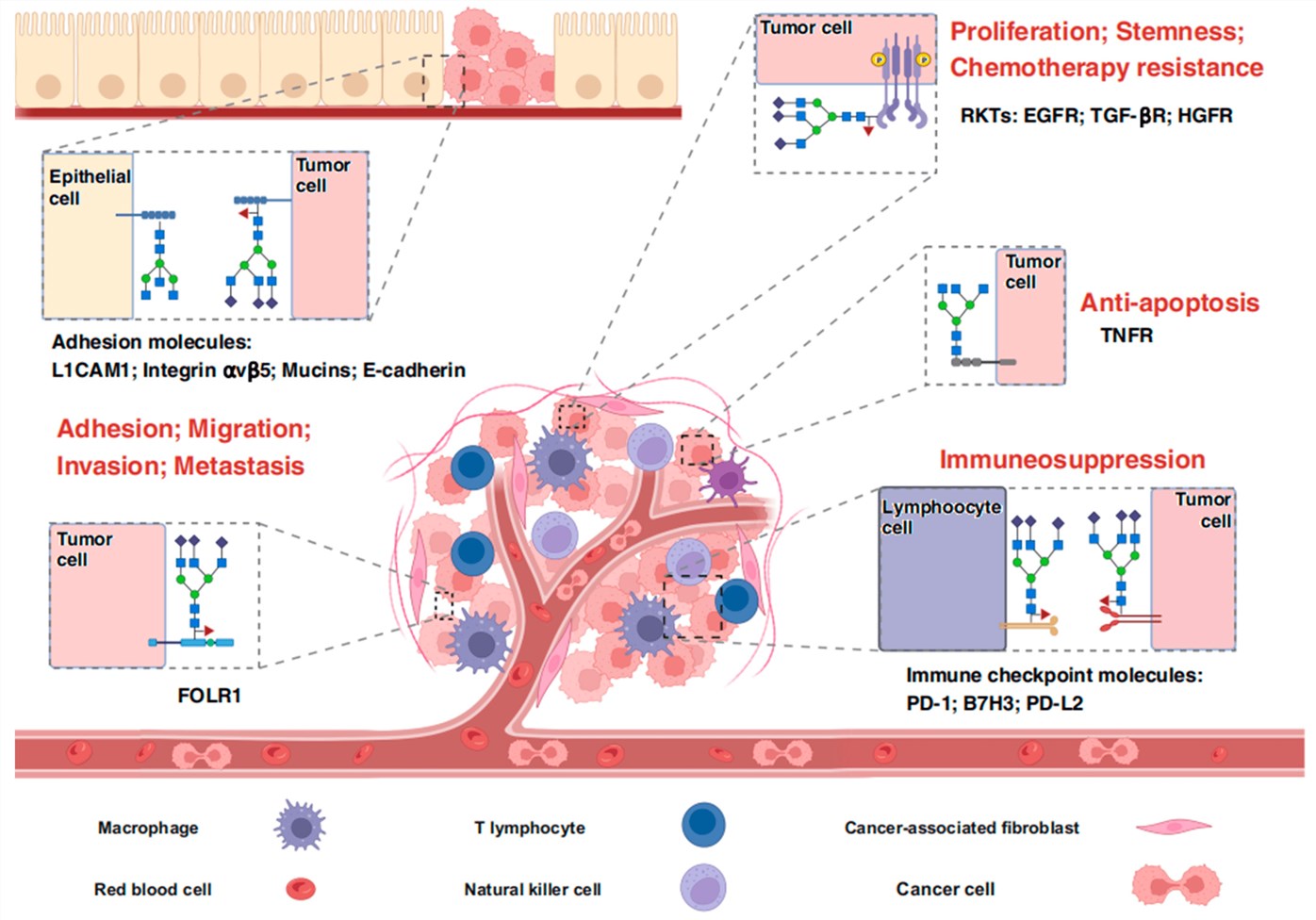 Fig.1
Proteins interacting with FUT8 and their biological effects in tumor progression.1
Fig.1
Proteins interacting with FUT8 and their biological effects in tumor progression.1
FAQs
How do you ensure your anti-FUT8 antibodies do not cross-react with other FUT family members like FUT4 or FUT9?
Specificity is achieved through rigorous antigen design. FUT8 belongs to Clade 1, while FUT4 and FUT9 belong to Clade 2, meaning they have significant structural differences. We select peptide sequences from the unique SH3 domain or specific loop regions of FUT8 that are absent in other FUTs. Furthermore, we employ negative absorption against recombinant FUT4/9 proteins to deplete any cross-reactive antibodies.
Can these antibodies be used for detecting FUT8 in serum samples for diagnostic research?
Yes, but this typically requires a sandwich ELISA format using a matched pair of monoclonal antibodies. Our service can be tailored to generate such pairs. While FUT8 is primarily an intracellular enzyme, soluble forms can be shed into the serum. Our antibodies are developed to recognize epitopes that are preserved in this soluble form, making them suitable for developing serological assays for research use.
Do you offer antibodies against the novel POFUT3 and POFUT4 enzymes?
Yes. We recognize the emerging importance of POFUT3 (formerly FUT10) and POFUT4 (formerly FUT11) in modifying EMI domains. We can design immunogens based on the predicted catalytic domains of these enzymes to generate antibodies that distinguish them from the structurally related POFUT2 (which modifies TSRs).
Can your antibodies be used to validate FUT inhibitors like SGN-2FF?
Absolutely. Our antibodies are valuable tools for pharmacodynamic studies. They can be used to measure the expression levels of the FUT enzymes themselves (to check for compensatory upregulation) or, in conjunction with anti-glycan antibodies, to assess the functional readout of inhibition (e.g., reduced Core Fucose levels after SGN-2FF treatment).
What is the typical timeline for a custom rabbit polyclonal antibody project?
A standard rabbit polyclonal antibody project takes approximately 70–80 days. This includes 2 weeks for peptide synthesis and conjugation, 6–8 weeks for the immunization protocol (multiple boosts), and 1–2 weeks for purification and initial ELISA validation. We provide interim reports after test bleeds so you can monitor the titer development.
Do you offer validation in specific cancer cell lines?
Absolutely. We can validate the generated antibodies using Western Blotting or Immunocytochemistry (ICC) on a panel of relevant cell lines. For gastric cancer research, we commonly use lines such as MKN45, AGS, or SGC-7901. We can also compare signal intensity between wild-type and FUT8-knockout cells to provide definitive proof of specificity.
Are your services for clinical diagnostic use?
No. All services and products provided by Creative Biolabs, including our custom antibody development, are strictly for Research Use Only (RUO). They are not intended for use in diagnostic or therapeutic procedures in humans or animals. However, the data generated using our tools can form the basis for your subsequent translational research and preclinical studies.
What Our Customers Say
"We were struggling to find a commercial antibody that could reliably detect FUT8 in our gastric cancer tissue arrays without high background. The polyclonal antibody developed by Creative Biolabs was clean and specific. It allowed us to clearly visualize the overexpression in tumor versus normal tissue."
"The project management was excellent. We needed an antibody for a sandwich ELISA setup to detect serum FUT8. They suggested a monoclonal pair development strategy. The resulting sensitivity was impressive, reaching the pg/mL range we needed for our pilot study."
"Specificity was our main concern due to the homology with FUT4. Creative Biolabs performed a thorough negative selection during purification. The final antibody showed no cross-reactivity in our Western Blot controls. Highly recommended for enzyme target projects."
"We utilized their service to generate an anti-FUT8 antibody for checking knockdown efficiency in our gene-edited cell lines. The data was clean and reproducible, which was crucial for our publication."
Reference:
- Shi, M., Nan, X.R., and Liu, B.Q. "The multifaceted role of FUT8 in tumorigenesis: from pathways to potential clinical applications." International Journal of Molecular Sciences 25.2 (2024): 1068. Distributed under Open Access license CC BY 4.0. https://doi.org/10.3390/ijms25021068.
Supports
- Glycosyltransferase & Glycosidase Substrate Microarray
- Glycosylation Analysis
- Custom Glycosylation of Biomolecules