Anti-GlcNAc-transferase (GnT) Antibody Development Service
N-acetylglucosaminyltransferases (GnTs) are a pivotal family of glycosyltransferases responsible for the branching of N-linked glycans. These enzymes, encoded by the MGAT gene family, initiate the formation of complex and hybrid N-glycans by transferring N-acetylglucosamine (GlcNAc) from UDP-GlcNAc to the mannose core of growing oligosaccharides. The branching patterns dictated by GnTs—specifically MGAT1, MGAT2, MGAT3, MGAT4, and MGAT5—serve as molecular switches that regulate cell surface receptor stability, half-life, and interaction with the extracellular matrix. Aberrant expression of these enzymes is frequently observed in pathological conditions, particularly in the acquisition of metastatic potential in cancer cells. Creative Biolabs leverages decades of expertise in glycobiology to provide a comprehensive Anti-Glycan Related Enzyme Antibody Development Service. We specialize in generating high-affinity antibodies against specific GnT isoforms, enabling researchers to dissect the complex biosynthetic pathways of N-glycosylation with precision.
Understanding GlcNAc-transferases (GnTs) and Their Biological Roles
The structural diversity of N-glycans is primarily determined by the specific GlcNAc-transferases that act in the Golgi apparatus. Each enzyme in this family has a unique substrate specificity and creates a distinct linkage, influencing the biological function of the final glycoprotein.
| Enzyme | Gene | Enzymatic Action & Linkage | Biological Significance |
|---|---|---|---|
| GnT-I | MGAT1 | Adds β1-2 GlcNAc to the α1-3 mannose arm. | Essential for the conversion of high-mannose to hybrid/complex glycans. Knockout is embryonically lethal. |
| GnT-II | MGAT2 | Adds β1-2 GlcNAc to the α1-6 mannose arm. | Required for complex N-glycan synthesis. Deficiency leads to CDG type IIa. |
| GnT-III | MGAT3 | Adds "bisecting" β1-4 GlcNAc to the core β-mannose. | Suppresses further branching (antagonizes GnT-IV/V). Linked to tumor suppression and ADCC modulation. |
| GnT-IV | MGAT4 | Adds β1-4 GlcNAc to the α1-3 mannose arm. | Regulates glucose transporter (GLUT2) retention and pancreatic beta-cell function. |
| GnT-V | MGAT5 | Adds β1-6 GlcNAc to the α1-6 mannose arm. | Creates β1-6 branching, a hallmark of cancer metastasis. Enhances lattice formation and receptor signaling. |
Why is Developing Anti-GnT Antibodies Challenging?
Despite their biological importance, commercially available antibodies against GnTs often lack the specificity or sensitivity required for rigorous biochemical analysis. Developing high-quality reagents for this target class presents several unique obstacles:
Sequence Homology & Conservation
The catalytic domains of various glycosyltransferases, including the MGAT family and related enzymes like B3GNT, often share conserved structural motifs. This high degree of sequence similarity can lead to cross-reactivity, where an antibody raised against MGAT1 might inadvertently recognize MGAT2 or other family members, confounding data interpretation.
Low Immunogenicity & Expression
As Golgi-resident type II transmembrane proteins, GnTs are often expressed at low levels relative to structural proteins. Furthermore, the active sites of these enzymes are evolutionarily conserved, which can result in low immunogenicity in host animals. Standard immunization protocols frequently fail to elicit a high-titer response against the native conformation of the enzyme.
Protein Instability
Purifying full-length, active glycosyltransferases for use as immunogens is notoriously difficult. These proteins are often unstable when extracted from the membrane environment, prone to aggregation, and may lose the conformational epitopes required for generating antibodies that work in immunoprecipitation or flow cytometry.
Lack of Standardization
Many commercially available anti-glycosyltransferase antibodies are validated only by Western Blot using overexpression lysates. They often fail in endogenous settings or more demanding applications like immunohistochemistry (IHC), where specific localization to the Golgi apparatus is critical for verification.
Our Custom Anti-GnT Antibody Development Services
Creative Biolabs overcomes these hurdles through a tailored development pipeline designed specifically for carbohydrate active enzyme antibodies. We employ a multi-faceted approach to ensure the production of antibodies that are both highly specific to the target isoform and sensitive enough to detect endogenous expression levels.
Antigen Design & Preparation Strategy
We do not rely on generic peptide synthesis. Our team carefully analyzes the crystal structure and homology models of the target GnT.
- Specific Peptide Selection: We identify surface-exposed, isoform-specific loops that are distinct between MGAT1, MGAT2, and MGAT5 to avoid cross-reactivity.
- Recombinant Protein Expression: For applications requiring recognition of conformational epitopes, we express the soluble catalytic domain in mammalian systems (HEK293 or CHO) to ensure proper post-translational modifications.
- DNA Immunization: To target the native protein structure, we utilize genetic immunization techniques, bypassing the need for protein purification and ensuring the host is exposed to the antigen in its native context.
Host Species & Antibody Formats
We offer flexibility in host selection to match your downstream application needs.
- Rabbit Monoclonal Antibodies (RabMAb): Ideal for IHC and detecting low-abundance targets due to the rabbit's superior immune system diversity and high affinity.
- Mouse Monoclonal Antibodies: The gold standard for assay development and reliable, long-term supply.
- Polyclonal Antibodies: Available in rabbit, goat, or chicken, providing robust signal amplification for Western Blotting.
- VHH (Single Domain) Antibodies: Derived from llama/alpaca, these small antibodies can access cryptic clefts in the enzyme active site, potentially acting as neutralizing or inhibitory antibodies.
Rigorous Screening & Validation
A generated antibody is only as good as its validation. We employ a "negative selection" strategy during screening.
- Counter-Screening: Candidates are screened against closely related homologs (e.g., screening an MGAT antibody against MGAT1, MGAT2, and MGAT4 simultaneously) to eliminate cross-binders.
- Application-Specific Testing: We validate clones in the specific format you intend to use, whether it is Western Blot, IHC on tissue arrays, or Immunofluorescence (IF) for Golgi co-localization studies.
- Knockout/Knockdown Verification: Whenever possible, we validate specificity using knockout or siRNA knockdown cell lines to confirm target specificity.
Why Choose Creative Biolabs?
Expert Antigen Design
We utilize structural biology insights to select epitopes that guarantee isoform specificity.
Validated Performance
Our antibodies are tested in real biological contexts, ensuring they work on endogenous proteins.
Broad Enzyme Coverage
From MGAT antibody development to sialyltransferase antibody and fucosyltransferase antibody targets.
Rapid Turnaround
Streamlined workflows allow us to deliver purified, validated antibodies in as little as 12-16 weeks.
Start Your Custom GnT Antibody Project
Published Data
N-acetylglucosaminyltransferase V (GnT-V), a pivotal glycosyltransferase responsible for catalyzing the formation of β1,6-GlcNAc branches on N-linked glycans, is increasingly recognized for its role in pathological tissue remodeling beyond its well-known involvement in oncology. In a comprehensive 2024 investigation, researchers utilized specific antibodies to explore the involvement of GnT-V in the pathogenesis of diabetic cardiomyopathy (DCM). Through precise immunohistochemical and immunofluorescence analyses of cardiac tissue sections, the study revealed a marked upregulation of GnT-V protein expression in diabetic models compared to healthy controls. This aberrant glycosylation pattern was strongly localized to cardiomyocytes and fibroblasts, correlating with maladaptive cardiac remodeling.
Further functional validation involved the targeted silencing of GnT-V using adeno-associated viral vectors. The antibody-based characterization of tissue samples demonstrated that reducing GnT-V levels significantly alleviated myocardial hypertrophy, evidenced by normalized cardiomyocyte size and reduced expression of hypertrophic markers such as ANP and β-MHC. Moreover, the intervention effectively suppressed interstitial fibrosis and collagen deposition via the modulation of integrin β1 and TGF-β1 signaling pathways. These data, derived from high-quality immunodetection assays, underscore the utility of anti-GnT-V antibodies in dissecting complex glyco-pathologies and identifying novel therapeutic avenues for preventing heart failure in diabetic conditions.
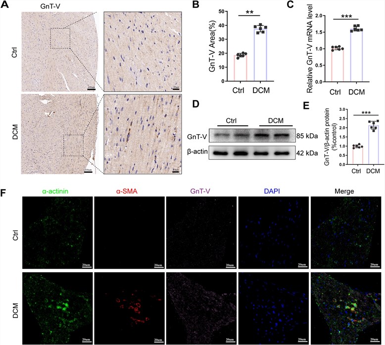 Fig.1
Immunohistochemical analysis of GnT-V expression in diabetic cardiac tissues. The antibody specifically detects endogenous GnT-V upregulation (brown staining) in the diabetic myocardium compared to control tissues.1
Fig.1
Immunohistochemical analysis of GnT-V expression in diabetic cardiac tissues. The antibody specifically detects endogenous GnT-V upregulation (brown staining) in the diabetic myocardium compared to control tissues.1
FAQs
How do you ensure your anti-GnT antibodies do not cross-react with other glycosyltransferases?
Specificity is our top priority. We begin by aligning the protein sequences of the target GnT (e.g., MGAT5) with its closest homologs (e.g., MGAT1, MGAT2, MGAT4). We design immunogens based on unique regions, typically loops distant from the conserved catalytic pocket. During screening, we use counter-selection ELISA or protein arrays containing these related enzymes to discard any cross-reactive clones immediately.
Can you develop antibodies that distinguish between active and inactive forms of the enzyme?
This is challenging but possible. If the active and inactive forms have distinct conformational differences (e.g., phosphorylation states or proteolytic cleavage products), we can design strategies to target these specific features. However, for most standard applications, we focus on detecting total protein levels with high specificity.
Do you offer services for other glycan-related enzymes besides GnTs?
Yes, our Anti-Glycan Related Enzyme Antibody Development platform is comprehensive. We can generate antibodies against a wide range of targets, including fucosyltransferase antibody targets (FUT8, FUT4), sialyltransferase antibody targets (ST6GAL1, ST3GAL), sialidase antibody targets (NEU1, NEU3), and other enzymes like heparanase or OGT (O-GlcNAc transferase).
What is the typical timeline for a custom monoclonal project?
A standard mouse monoclonal antibody project typically takes about 4-6 months from antigen preparation to delivery of purified hybridoma supernatant. This includes immunization, fusion, screening, and subcloning. Expedited services or recombinant antibody approaches can sometimes shorten this timeline.
Can these antibodies be used for flow cytometry to detect intracellular GnTs?
Yes, but cell permeabilization is required since GnTs are located in the Golgi apparatus. We can include flow cytometry screening on permeabilized cells as part of the validation package to ensure the antibody works in this application.
What Our Customers Say
"Finding a good MGAT5 antibody has been a nightmare for our lab. The commercial ones were essentially garbage for IHC. Creative Biolabs designed a custom rabbit mAb for us targeting a specific loop, and the difference is night and day. Clean Golgi staining, zero background."
"We needed to differentiate between MGAT1 and MGAT2 in a knockout model. Their team suggested a peptide immunization strategy that worked perfectly. The validation data they provided gave us total confidence before we even received the vial."
"Excellent service for generating antibodies against difficult enzymes. We used their service for a B3GNT target and the yield and purity were impressive. The communication was professional throughout the project."
"The project manager really understood the nuances of glycosyltransferases. They didn't just sell us a product; they helped design the antigen to avoid the catalytic domain which is too conserved. Highly recommended."
Reference:
- Zhao, R., et al. "Inhibition of N-acetylglucosaminyltransferase V alleviates diabetic cardiomyopathy in mice by attenuating cardiac hypertrophy and fibrosis." Nutrition & Metabolism 21.1 (2024): 53. Distributed under Open Access license CC BY 4.0, without modification. https://doi.org/10.1186/s12986-024-00797-w
Supports
- Custom Glycosylation of Biomolecules
- Glycosylation Analysis
- Glycosyltransferase & Glycosidase Substrate Microarray