Creative Biolabs works with researchers who need anti-GT1b antibodies that remain
informative beyond a single purified-antigen assay. GT1b is displayed in neuronal membranes, where
glycan orientation, lipid packing, surface density, and neighboring gangliosides can all influence
antibody recognition. We bring these variables into the project discussion from the beginning, then
align antigen design, discovery, specificity screening, and application-focused validation with your
neurological research model.
GT1b shares substantial structure with GD1a, GD1b, GT1a, and GQ1b. A useful reagent therefore needs a defined
cross-reactivity profile rather than a target-only binding result. We provide an end-to-end custom
anti-ganglioside antibody service dedicated to generating high-quality anti-ganglioside antibodies with
precise specificity to GT1b ganglioside. Built by senior scientists with decades of experience in glycan
immunology and membrane biochemistry, our platform integrates rational antigen design for glycolipids,
multi-modal discovery platforms, glycan-focused screening, and rigorous QC. We are committed to offering
reproducible anti-GT1b antibody candidates you can trust for neurological autoimmunity research. Off-the-shelf anti-GT1b
antibodies made for ELISA, IF, IHC, and WB are also provided if you require a fast start.
Challenges in Developing GT1b-Specific Antibodies
GT1b presents a dense, highly sialylated headgroup and shares
important structural features with GD1a, GD1b, GT1a, and GQ1b. That combination can produce weak immune
responses, broad polysialic-acid recognition, or binding that changes when the antigen moves from a coated plate
to a membrane. A useful development plan therefore begins by defining which related gangliosides must be
rejected and which presentation best reflects the final assay.
Identify gangliosides that must be excluded and any complexes that should be recognized.
Specify species, neural model, fixation state, and intended readout.
Define whether high avidity, monovalent binding, or functional perturbation is the
priority.
Our Anti-GT1b Antibody Development Services
Our current GT1b service page describes a flexible program
spanning strategic antigen design, hybridoma or display-based discovery, glycan-focused specificity screening,
application validation, and optional recombinant formatting. Rather than treating these as a fixed package,
Creative Biolabs helps you select the elements that answer your research question and fit the antibody format
you ultimately need.
GT1b Immunogen and Antibody Development
GT1b may be presented through a defined conjugate, liposome, or another membrane-mimetic system. Antigen
identity, lipid composition, surface density, linker exposure, and carrier controls are considered together
to reduce selection of presentation artifacts.
Specificity and Membrane-Context Validation
Specificity assessment compares GT1b with structurally adjacent gangliosides and then tests whether the
selected signal is retained in liposomal, cellular, or tissue context. Array selectivity alone is not
treated as proof of biological-context performance.
Preserving GT1b Presentation in Antigen Design
Soluble or plate-adsorbed ganglioside can adopt presentations
unlike a lipid bilayer. Carrier conjugates, liposomes, or other membrane mimetics can expose the glycan
headgroup while retaining a hydrophobic environment. Parallel formats help distinguish genuine GT1b recognition
from coating or lipid-phase artifacts.
Presentation
Use
Guardrail
Carrier conjugate
Immune focusing
Reject carrier and linker binding
Liposome
Membrane-like display
Control lipid composition and surface density
Defined array
Specificity mapping
Confirm hits in a second format
Cells/tissues
Biological relevance
Use ganglioside-status and enzymatic controls
The screening cascade is most informative when neural-cell
accessibility is assessed alongside ganglioside complexes. One readout describes structural preference; the
other tests whether the preference survives the valency, accessibility, and competing surfaces present in the
intended model. Candidates that change rank between these settings are not automatically unsuitable, but the
difference must be explained before a lead is selected for an application-specific study.
Our Development Workflow
The workflow moves from controlled structural questions to
native-context confirmation. We first agree on the GT1b presentation and counter-target panel, generate and
screen candidates, then compare shortlisted binders across related gangliosides and a membrane-relevant format.
Only candidates with an interpretable recognition profile are carried into the requested cell, tissue, or
downstream engineering work.
01
Antigen Design
02
Screening
03
Profiling
04
Validation
Project Requirements and Deliverables
Project Input
Project Requirements
GT1b source, composition, purity, and planned presentation format
GQ1b, GD1a, GD1b, GT1a, or other required counter-targets
Cell, tissue, liposome, or purified-ganglioside sample context
Desired antibody format and intended research readout
Project Output
Deliverables and Validation Scope
Depending on the agreed scope, outputs may include antibody
candidates, ganglioside-panel screening data, and membrane-context validation results. Interpretation
distinguishes structural selectivity from assay- or avidity-dependent signal.
Project inputs should describe fixation sensitivity in enough
detail to reproduce the intended antigen context and should identify how regional tissue distribution will be
evaluated. This information affects reagent choice, control construction, and the meaning of a negative result.
If a customer sample is the final destination, its preparation history and expected target abundance are
considered during planning rather than introduced only after purified-antigen screening is complete.
Specificity Mapping Across Related Gangliosides
Screen target binding in at least two GT1b presentations.
Compare GD1a, GD1b, GT1a, GQ1b, and other study-relevant gangliosides.
Use asialo and lipid-only controls to detect charge- or membrane-driven binding.
Re-rank candidates under the final assay geometry before format engineering.
Data interpretation distinguishes a measured interaction
profile from a universal property of the antibody. Evidence for membrane-mimetic validation is limited to the
structures, concentrations, and presentation formats actually tested. Changes in density, matrix, or antibody
format can alter apparent selectivity through avidity. Reporting these boundaries makes it easier to decide
whether additional controls, a second assay geometry, or a different clone is needed.
Research Applications
Profile GT1b and related ganglioside recognition in antibody panels.
Map membrane or tissue distribution in research models.
Investigate GT1b-dependent receptor complexes and neurite-outgrowth mechanisms.
Develop research capture, competition, or imaging reagents.
Clinical interpretation of patient antibodies is outside the
scope of this research-use service.
For research applications, functional perturbation is evaluated
as a separate question from antigen recognition. Binding may justify advancing a clone, but it does not by
itself demonstrate blocking, internalization, signaling, staining, or biological activity. Those endpoints
require their own controls and model-specific acceptance logic. Maintaining this separation prevents a
descriptive interaction reagent from being overinterpreted as a functional antibody.
Recommended Services or Products You May Need
The final clone discussion integrates structural data,
orthogonal confirmation, and sample relevance rather than relying on a single highest signal. Trade-offs among
GT1b surface density, ganglioside complexes, and antibody avidity are reviewed against the stated research
objective. This approach allows a narrowly selective binder and a deliberately broader binder to be evaluated on
their intended merits, provided that the recognition envelope and its limitations are documented clearly.
In addition to our custom development services, Creative
Biolabs offers a collection of ready-to-use anti-GT1b
antibodies with verified specificity and performance. These off-the-shelf reagents are ideal for ELISA,
IF, IHC, WB and other research applications targeting GT1b ganglioside. Each product is rigorously tested and
accompanied by detailed datasheets to support your experimental workflows with confidence and convenience.
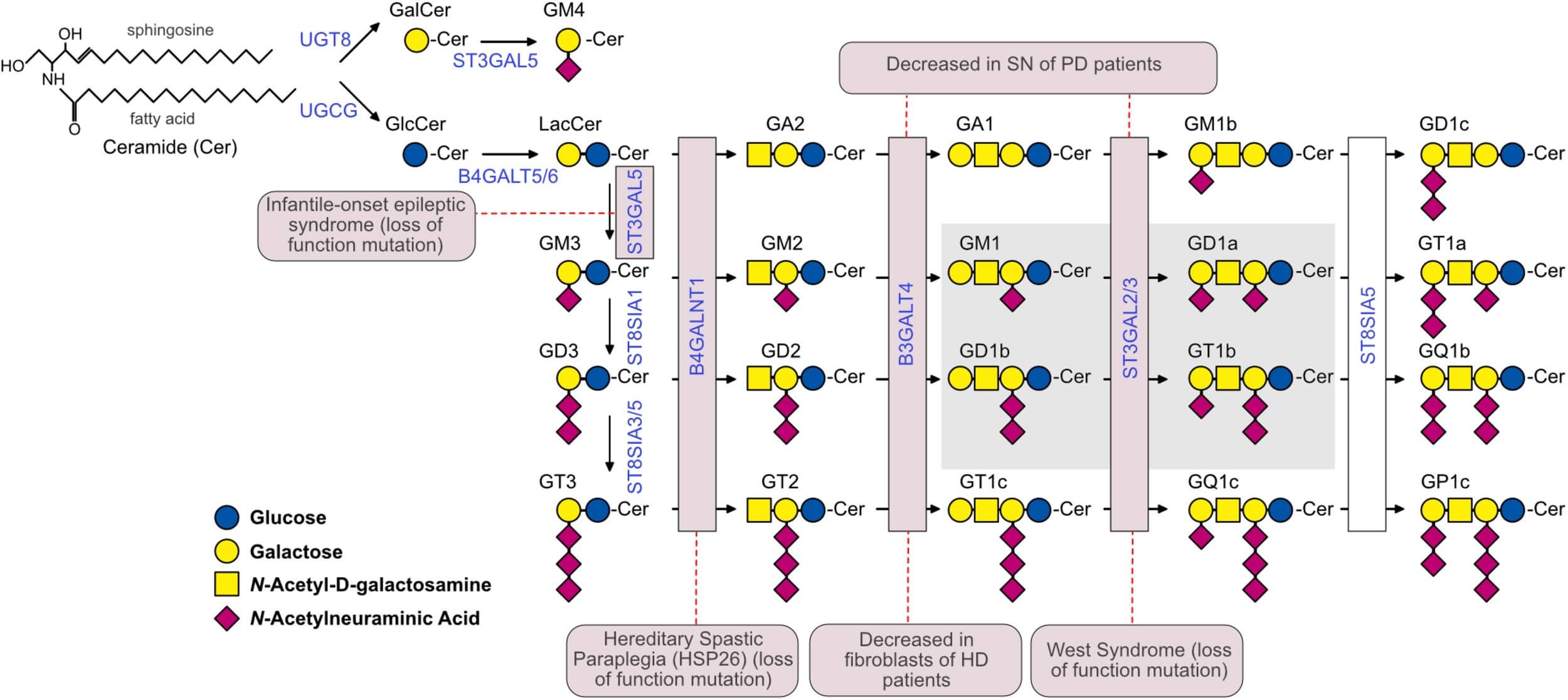
Sipione and colleagues place GT1b within the shared biosynthetic network of major brain gangliosides and
describe how ganglioside ceramide anchors position glycan headgroups at the outer membrane leaflet. Read
together, these features support two practical selection decisions: use membrane-aware antigen formats to
evaluate presentation-dependent binding, and include biosynthetically or structurally related gangliosides
as counter-targets when defining an anti-GT1b recognition profile.
Fig.1 GT1b within the brain ganglioside biosynthetic network and its relationship to structurally
related gangliosides.1
Plan Your Anti-GT1b Antibody Project
Share the GT1b source and presentation you plan to use, the gangliosides that must be excluded, your
biological model, intended assay, and preferred antibody format. If you already have an antigen or candidate
antibody, include its composition, handling information, and available data. Creative Biolabs will use these
details to discuss a focused research plan.
Discuss Your Anti-GT1b
Project
Frequently Asked Questions
Why is GT1b more difficult to target than a typical protein antigen?
GT1b is a membrane-embedded glycolipid with a highly
sialylated carbohydrate headgroup. Its immunogenicity and apparent epitope can change with carrier choice,
lipid environment, surface density, and assay geometry. Antibody development must therefore coordinate antigen
presentation with related-ganglioside screening instead of relying on a conventional protein workflow.
Which gangliosides should be included in an anti-GT1b specificity panel?
The panel is usually built around structurally or biologically
relevant neighbors such as GD1a, GD1b, GT1a, GQ1b, and other gangliosides present in the study system. The
final selection should reflect the intended sample and research question, because an unnecessarily broad panel
may add work without improving interpretation.
Should GT1b be presented as a conjugate or in liposomes?
Both formats can be useful, but they answer different
questions. A conjugate offers controlled screening and may improve immunogenicity, whereas liposomes provide a
membrane-like environment for the glycolipid. Many projects benefit from using one format for discovery and
another for confirmation, provided suitable carrier and lipid controls are included.
Can customer-supplied GT1b be used in the project?
Customer-supplied material can be reviewed for compatibility
with the proposed immunization or screening route. Please provide composition, purity information, solvent,
concentration, storage history, and handling requirements. These details help determine whether the material
can be incorporated directly or whether an alternative presentation should be prepared for consistent testing.
When should a GT1b antibody be tested on cells or tissues?
Cell or tissue testing becomes important when the final
application depends on native accessibility, local density, fixation, or surrounding membrane components. It
is generally most efficient after primary binding and counter-screening have narrowed the candidate set.
Matched positive and negative models are needed to interpret a weak or absent signal responsibly.
Can an anti-GT1b lead be reformatted or labeled?
Sequence-defined leads may be considered for IgG, Fab, scFv,
single-domain, or labeled research formats according to project scope. Reformatting can change valency,
avidity, and access to membrane-displayed GT1b, so the new construct should be reconfirmed against the target,
relevant counter-targets, and the intended application rather than accepted by sequence alone.
References
Sipione, Simonetta, et al. "Gangliosides in the Brain: Physiology, Pathophysiology and Therapeutic
Applications." Frontiers in Neuroscience 14 (2020): 572965. Distributed under Open Access
license CC BY 4.0, without modification. https://doi.org/10.3389/fnins.2020.572965
Project Inquiry
!For Research Use
Only.Not for clinical or diagnostic use.