The affinity of antibodies obtained from hybridomas often falls short of the level of effective clinical use, in part because of the in vivo affinity ceiling, and thus improving antibody affinity is often required. In the last decade, scientists of Creative Biolabs have made considerable efforts to develop mammalian cell surface display technology. With our extensive experience, we now offer antibody affinity maturation services using mammalian cell display technology for our worldwide customers.
Generating high-affinity antibodies to important biomolecules for clinical trials is a critical and challenging task. The affinity of antibodies obtained from hybridomas often falls short of the level of effective clinical use, partly because of the in vivo affinity ceiling, and thus improving antibody affinity is often required. Mammalian cell display is an effective method for isolating whole IgG and scFv with high affinity and other specific biological functions. After constructing and screening a comprehensive mammalian cell display library, appropriate mutagenesis strategies are essential to increase antibody affinity to a satisfactory level. Mammalian cell display relies on the transient transfection of antibody-encoded DNA to promote very high levels of antibody expression in mammalian cells.
There are a variety of methods, ranging from random mutagenesis, site-directed mutagenesis in CDR and framework regions, to in vitro somatic hypermutation (SHM), resulting in significant optimization of antibody affinity, stability, and other characteristics. We have used mammalian cells to display single-chain antibody fragments (scFv) for affinity maturation. Mammalian cell display is used to isolate and engineer scFv, Fab or whole IgG to increase affinity and other specific biological functions. Flow cytometry increases the sensitivity of the screening, allowing us to isolate high-affinity antibodies.
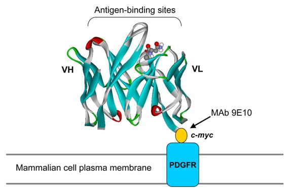
 Fig.1 Schematic illustration of surface display on mammalian cells. (Ho, 2009)
Our strategy includes suitable transient expression system and appropriate target protein selection. The detection of cells bound to target proteins is done by FACS. Cells that bind to the target protein are detected by FACS. Cells transfected with the scFv library are sorted by flow cytometry and only cells in which the scFv binds to the target protein are collected. Plasmid DNA is isolated from these cells and amplified in bacteria. These scFv DNAs are further enriched until the scFv with increased affinity for the target protein can be isolated.
With our extensive knowledge and experience, we can provide global customers with the most suitable technical services to meet your research and project goals. For more detailed information, please feel free to contact us or directly sent us an inquiry.
Reference
All listed services and products are For Research Use Only. Do Not use in any diagnostic or therapeutic applications.