Introduction of Complement System
Complement system was discovered by Jules Bordet as the heat-labile component of normal plasma that results in the opsonization and killing of bacteria. Complement system is one of the first lines of defense in innate immunity and is crucial for cellular integrity, tissue homeostasis and modifying the adaptive immune response. The activated complement system directly initiates the immune effector functions and modulates the intensity of the response in a self-controlling manner. This allows the appropriate innate immune response, which is needed for recognition and removal of infectious agents or modified self-cells, shown in Fig.1. These complement reactions lead to a moderate and controlled outcome of complement activation, which is beneficial for the host but detrimental for the invading microorganism.
Complement Component/Proteins
There are a group of soluble plasma proteins in the complement system that interact with one another in two distinct enzymatic activation cascades (the classical and alternative pathways) and in the nonenzymatic assembly of a cytolytic complex (the membrane attack complex pathway, also called the terminal pathway), shown in Fig.1 and Fig.2. The third activation pathway is termed the lectin pathway. The component proteins are shown in Fig. 2. Enzymatic cleavages are represented by thick arrows. The lectin pathway differs from the classical pathway only in that C1 complex is replaced by the MBP-MASP complex. Regulators act to inhibit either the enzymes of the activation pathways (activated C1, C3 convertases, and C5 convertases) or assembly of the MAC.
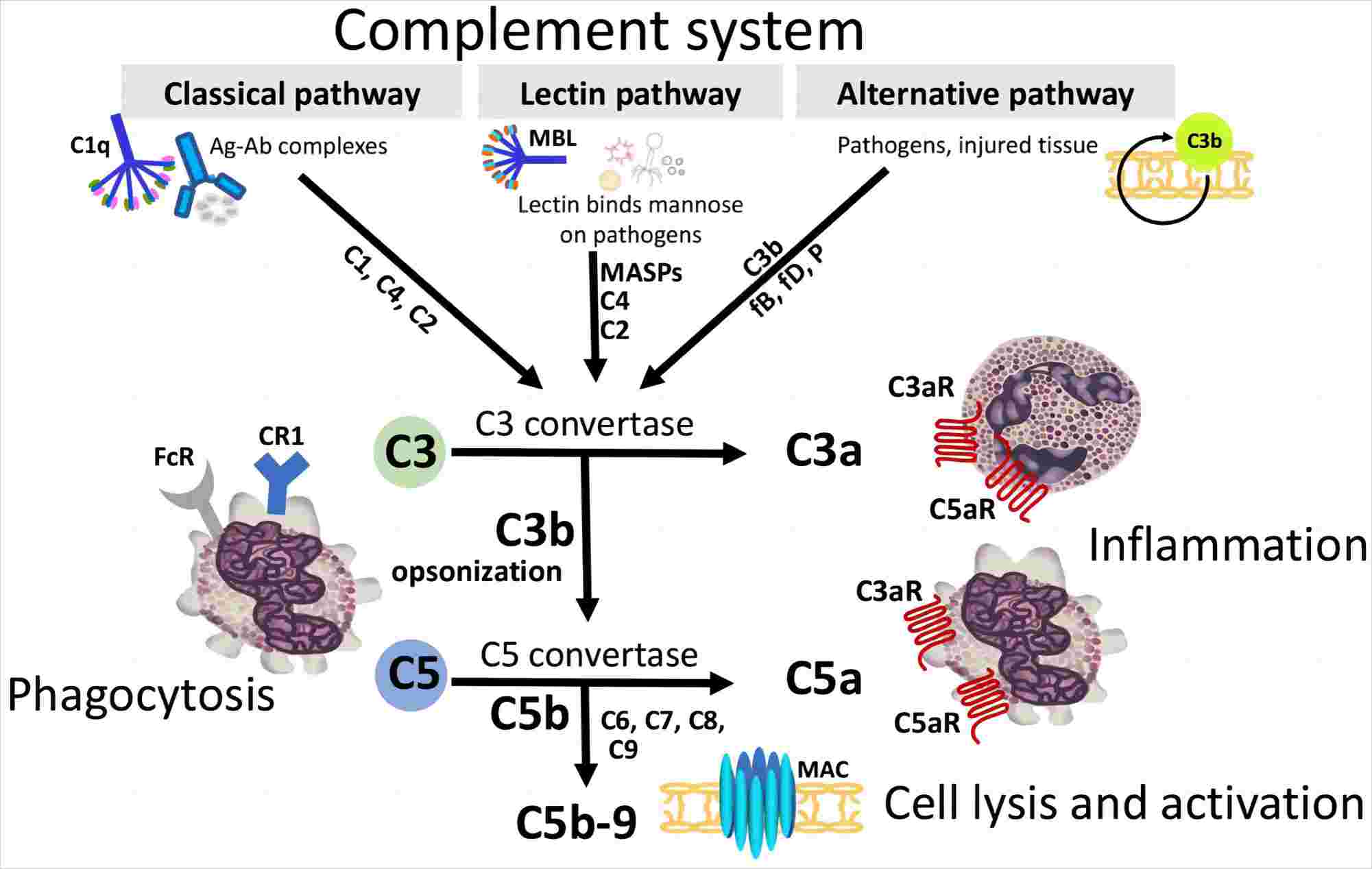
Fig. 1 The complement system and its control.1, 2
Complement Component/Protein Structure
The complement component/proteins with their structure are described below in Table 1.
Table.1 The Component Proteins of the Complement System.
|
Component
|
Structure
|
|
Classical pathway
|
C1
|
Complicated molecule, composed of 3 subunits, C1q (460 kDa), C1r (80 kDa), C1s (80 kDa) in a complex (C1qr2s2)
|
|
C4
|
3 chains (α, 97 kDa; β, 75 kDa, γ, 33 kDa); from a single precursor
|
|
C2
|
Single chain, 102 kDa
|
|
Alternative pathway
|
FB (Factor B)
|
Single chain, 93 kDa
|
|
FD (Factor D)
|
Single chain, 24 kDa
|
|
FP (Factor Properdin)
|
Oligomers of identical 53 kDa chains
|
|
C3
|
2 chains: α, 110 kDa, β, 75 kDa
|
|
Terminal pathway
|
C5
|
2 chains: 115 kDa, 75 kDa
|
|
C6
|
Single chain, 120 kDa
|
|
C7
|
Single chain, 110 kDa
|
|
C8
|
3 chains: α, 65 kDa, β, 65 kDa γ, 22 kDa
|
|
C9
|
Single chain, 69kDa
|
Functions of Complement Component Proteins
The complement system contains 12 soluble plasma proteins in the enzymatic activation cascades, including the classical, alternative, and lectin pathways. Most of the proteins are normally inactive. However, in response to the recognition of molecular components of microorganisms, they become sequentially activated in an enzyme cascade – the activation of one protein enzymatically cleaves and activates the next protein in the cascade. During these enzymatic cascades, various studies indicated that the control of these enzymatic cascades is provided by ten or more plasma and membrane-bound inhibitory proteins acting at multiple stages of the system and essential to prevent rapid consumption of the complement system in vivo. These proteins plays a central role in the complement system of the innate immune defense, which offers a system for the rapid destruction of a wide range of invading microorganisms.
Creative Biolabs offers a full range of complement component-related services and products, please do not hesitate to contact us for more details.
References
-
Girardi, Guillermina, et al. "Essential role of complement in pregnancy: from implantation to parturition and beyond." Frontiers in immunology 11 (2020): 1681.
-
under Open Access license CC BY 4.0, without modification
Related Product
For Research Use Only.
Related Sections: