Product List Background iC3b Functional Service
Background
iC3b is a fragment of complement component 3 (C3), consisting of two C3α' polypeptides that are disulfide-linked to the complete C3 β-chain. It plays a crucial role in the immune response by opsonizing pathogens and apoptotic cells, facilitating their clearance through phagocytosis by binding to complement receptors (CRs) like CR3. Elevated iC3b levels in the liquid phase are observed in various diseases, such as systemic lupus erythematosus, rheumatoid arthritis, sepsis, and myocardial infarction. This suggests its involvement in inflammatory processes and immune dysregulation, making it a potential biomarker and therapeutic target in related pathological conditions.
iC3b Formation
iC3b are generated by the cleavage of complement component C3b by factor I, facilitated by cofactors such as membrane cofactor protein (MCP) or CR1. Initially, C3b is generated through the cleavage of C3 by C3 convertase, resulting in the release of anaphylatoxin C3a and the deposition of C3b on target surfaces. Factor I then cleaves C3b at either one or two specific sites, forming iC3b. When factor I cleaves C3b at two sites, it produces a small fragment (C3f). Conversion of C3b to iC3b leads to functional changes in C3b, rendering iC3b less capable of participating in the amplification loop of complement activation and more focused on opsonization via CRs. Additionally, iC3b can undergo further cleavage to yield smaller fragments, C3c and C3dg, which maintain opsonic functions while potentially influencing immune regulation in various physiological and pathological contexts.
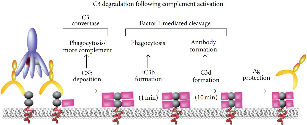 Fig.1 C3 degradation following complement activation.1
iC3b-Receptor Interaction
iC3b serves as an effective opsonin; it is recognized by CR2 and the two integrin receptors CR3 and CR4, all playing distinct roles in the immune response. CR2 is mainly expressed on B cells and promotes antigen presentation and B cell stimulation upon iC3b binding. CR3, composed of the integrin subunits αM and β2, exhibits high expression on the plasma membrane of myeloid cells such as dendritic cells, macrophages, and neutrophil granulocytes. Its presence can be enhanced by mobilizing it from storage granules upon cellular stimulation. It is crucial for the phagocytosis of iC3b-coated particles and modulates inflammatory responses. In addition, CR3 is involved in complement activation of the adaptive immune system. CR4 is found on macrophages, neutrophils, and monocytes, promoting phagocytosis and immune cell activation in response to iC3b-opsonized targets.
Antibodies Targeting iC3b
Antibodies targeting iC3b are pivotal for detecting and neutralizing complement activation products in various diseases. They are used in diagnostic assays to quantify iC3b levels, aiding disease monitoring and prognosis research. These antibodies also aim to neutralize iC3b-opsonized targets and modulate complement activation and inflammation by blocking iC3b interactions with its receptors, attenuating immune-mediated damage in conditions like autoimmune disorders and transplant rejection.
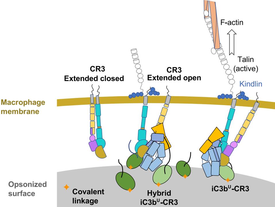 Fig.2 The iC3b-CR3 interaction across the gap between macrophage and iC3b- and C3dg-opsonized surface.2, 4
Creative Biolabs offers high-quality anti-iC3b antibody products for several applications, such as WB, FC, IHC, Neut, etc. In addition, highly specific human iC3b ELISA kits and native human complement iC3b protein are also available here.
iC3b Functional Service
Creative Biolabs offers a comprehensive suite of iC3b-related reagents, which include anti-iC3b antibodies, ELISA kits for iC3b detection, and human complement iC3b proteins. These tools are adept at identifying and scrutinizing the interactions involving the human iC3b protein and various other factors. Consequently, they serve as vital resources in research aimed at devising therapeutic strategies for disease management.
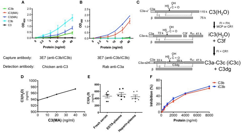 Fig.3 Distinctiveness of the C3(H2O) ELISA.3, 4
Researchers have rekindled interest in the alternative pathway activation of C3 due to the discovery of a C3(H2O) uptake route in human biospecimens. Historically, distinguishing C3(H2O) from C3 and its activation products posed challenges in human samples. Addressing these issues, a C3(H2O)-specific ELISA was developed to accurately measure C3(H2O) in human serum and plasma. To validate the assay’s specificity and assess interference from elevated C3b/iC3b levels, competition assays were conducted. Results indicated 50% inhibition of a 40 ng/ml C3(H2O) signal required 5500 ng/ml of C3b and 5000 ng/ml of iC3b. Therefore, at typical assay dilutions, interference by C3b or iC3b is minimal, ensuring precise C3(H2O) assessments.
Creative Biolabs offers specialized expertise in the functionality of iC3b through comprehensive analyses of interaction dynamics and thorough functional assessments. These meticulously designed services cater to the specific requirements of distinguished clients, significantly enhancing both clinical and scientific research endeavors.
References
-
Stowell, Sean R., et al. "Initiation and regulation of complement during hemolytic transfusion reactions." Journal of Immunology Research 2012.1 (2012): 307093. Distributed under Open Access license CC BY 3.0.
-
Fernández, Francisco J., et al. "The crystal structure of iC3b-CR3 αI reveals a modular recognition of the main opsonin iC3b by the CR3 integrin receptor." Nature communications 13.1 (2022): 1955.
-
Elvington, Michelle, et al. "Development and optimization of an ELISA to quantitate C3 (H 2 O) as a marker of human disease." Frontiers in immunology 10 (2019): 703.
-
Distributed under Open Access license CC BY 4.0, without modification.
 Datasheet