Product List Background Multiple myeloma cells (MM.1S) Aptamer Analysis
Background
Introduction of Multiple Myeloma Cells
Multiple myeloma is a malignancy originating from plasma cells, which are specialized white blood cells responsible for antibody production, alias myelomatosis. As a form of cancer, multiple myeloma is marked by the build-up of abnormal plasma cells in the bone marrow, resulting in tumors forming in various bones throughout the body. Healthy plasma cells produce antibodies essential for immune defense. However, in multiple myeloma, the excessive proliferation of malignant plasma cells results in the production of dysfunctional antibodies. This abnormal antibody production can lead to blood thickening and disrupt the bone marrow's ability to produce sufficient healthy blood cells. Furthermore, myeloma cells secrete factors that activate osteoclasts, contributing to bone damage and weakening. Additionally, kidney complications arise from the deposition of abnormal proteins produced by myeloma cells, which obstruct kidney tubules.
Multiple Myeloma Activating Signaling Pathways
The interactions of bone marrow microenvironment with the multiple myeloma cells trigger intracellular signaling through three distinct pathways:
1) First, soluble factors like cytokines and chemokines from accessory cells and osteoclasts, such as LIF and VEGF, stimulate multiple myeloma cell proliferation and progression from MGUS to multiple myeloma.
2) Second, physical interactions between multiple myeloma cells and other cellular components via adhesion molecules, like VCAM-1 and VLA-4, trigger proliferative and anti-apoptotic signals, activating the NF-κB pathway.
3) Third, exosomes from various cell types transfer bioactive molecules to multiple myeloma cells, activating survival pathways (c-Jun N-terminal kinase, p53, and Akt) and contributing to drug resistance.
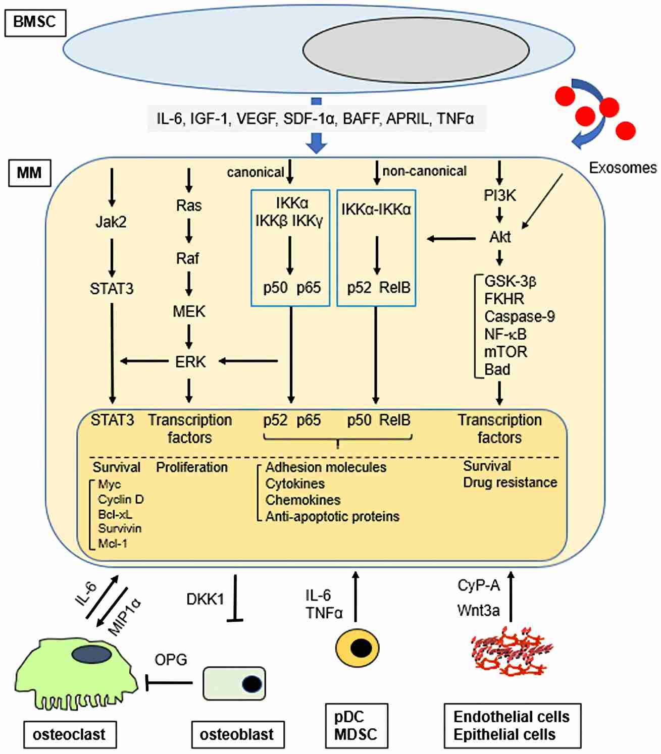 Fig. 1 Signaling pathway activation in multiple myeloma cells within the bone marrow microenvironment.1,4
Applications of Multiple Myeloma Cells Products
Multiple myeloma persists as an untreatable condition owing to its high recurrence rates and swift acquisition of drug resistance. The advent of mAbs offers targeted approaches with enhanced efficacy and reduces toxicity to multiple myeloma. For aptamer development, an ideal target would exhibit high and uniform expression in multiple myeloma cells, with minimal expression in normal cells to prevent off-target effects. Consequently, most aptamers under investigation target the same well-characterized molecules as antibodies, showing promising preclinical results.
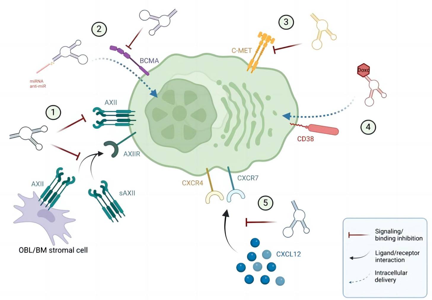 Fig.2 Aptamers for precision medicine in multiple myeloma.2,4
Targeting Annexin A2 in Multiple Myeloma: Aptamer-Based Therapeutic Approach
Annexin A2 (AXII), a calcium-dependent phospholipid-binding protein, is overexpressed in multiple myeloma plasma cell membranes and is inversely correlated with patient survival. AXII engages with its receptor AXIIR to bolster the adhesion and proliferation of multiple myeloma cells in the bone marrow milieu, fostering a tumor-supportive microenvironment. Targeting the AXII/AXIIR axis thus presents a promising therapeutic strategy. Researchers have isolated a single-stranded DNA aptamer that binds AXII with high affinity in the low nanomolar range, achieved through multiple rounds of protein-based SELEX. This aptamer specifically binds to AXII-expressing multiple myeloma cells in vitro and in vivo, inhibiting AXII-induced adhesion and progression, highlighting its potential for targeted treatment.
Multiple Myeloma Cells (MM.1S) Aptamer Analysis
Creative Biolabs provides a wide range of multiple myeloma cells (MM.1S) related products, including anti-multiple myeloma cells (MM.1S) aptamer. These products can effectively help to carry out your experiments and thus play an important role in your research.
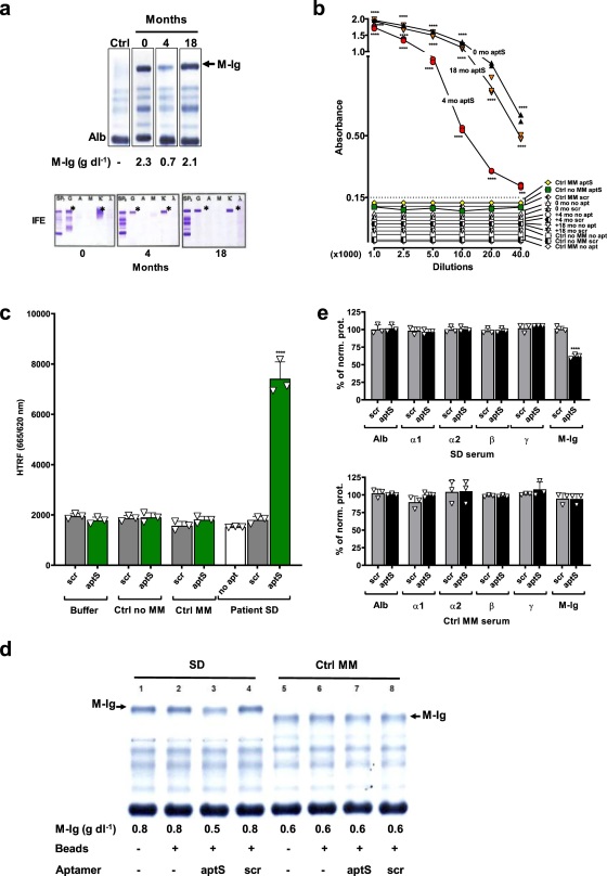 Fig. 3 Specificity and sensitivity of aptamer in patient serum.3,4
Minimal residual disease (MRD) is the main cause of relapse in multiple myeloma (MM), and how to identify it early is a key issue in treatment. Claudia Tapia-Alveal et al. reported a DNA aptamer generation method that can specifically bind to the patient's monoclonal immunoglobulin (M-Ig) Fab region, thereby quantitatively detecting MRD in serum.3,4 The overall selection strategy is to first eliminate binders that bind to common IgG, and then select the most sensitive target-specific aptamer in serum. Its advantages include automation, high sensitivity, and low sample consumption. The detection sensitivity is 2000 times higher than immunofixation and 20 times higher than mass spectrometry.
Creative Biolabs offers multiple myeloma cells (MM.1S) aptamer analysis, including SELEX service and other tailored functional services for our esteemed clients engaged in clinical and scientific research.
References
-
Hideshima, Teru, and Kenneth C. Anderson. "Signaling pathway mediating myeloma cell growth and survival." Cancers 13.2 (2021): 216.
-
Amundarain, Ane, et al. "Aptamers, a new therapeutic opportunity for the treatment of multiple myeloma." Cancers 14.21 (2022): 5471.
-
Tapia-Alveal, Claudia, Timothy R. Olsen, and Tilla S. Worgall. "Personalized immunoglobulin aptamers for detection of multiple myeloma minimal residual disease in serum." Communications Biology 3.1 (2020): 781.
-
Distributed under Open Access license CC BY 4.0, without modification.

 Datasheet