The in vitro use of polyclonal or monoclonal antibodies has greatly improved our ability to diagnose a number of diseases, including many cancers. As a leading company in the antibody development field, Creative Biolabs is offering first-class services of IVD antibody development for the diagnosis of diversified cancer types, such as brain cancer.
Introduction to Brain Cancer
Brain cancer refers to a diverse collection of cancers arising from different cells either within the brain (primary tumors) or from systemic tumors that have metastasized to the brain. The symptoms and signs of brain cancer are broad and nonspecific including headaches, weakness, seizures, vomiting and so on. To date, most brain tumors have no known cause, but risk factors that have been identified include inherited neurofibromatosis, exposure to vinyl chloride, Epstein–Barr virus, and ionizing radiation. Treatment of brain cancer usually includes some combination of surgery, radiation therapy, and chemotherapy. Different types of treatment are used depending on neoplasm type and grading, underlining the importance of a precise diagnosis.
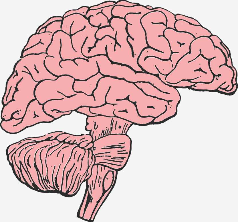
 Distributed under Pixabay License, from Pixabay.
Diagnosis of Brain Cancer
An accurate diagnosis of the type of the tumor is fundamental before a specific treatment can be started. When there is a suspicion of a brain tumor, the diagnostic procedure usually starts with a type of imaging technology such as an MRI scan or a CT scan, where neoplasms will often show as differently colored masses. Subsequently, the result needs to be confirmed and the tumor type and grade need to be determined by a neuropathologist using a tissue sample of the tumor obtained by biopsy or surgery. Brain cancer diagnosis made by conventional histological methods depends on the recognition of morphological patterns and the use of a number of special staining techniques. Although the majority of tumors of the central nervous system (CNS) can be diagnosed in this manner, there remains a significant proportion in which the diagnosis is uncertain and other ancillary techniques are necessary, including immunocytochemical staining and electron microscopy.
Immunohistological Staining Techniques
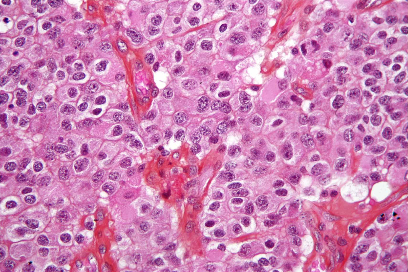
 Fig.1 An oligodendroglioma, H&E stain. Distributed under CC BY-SA 3.0, from Wiki, without modification.
Creative Biolabs offers affordable and reliable in vitro diagnostic (IVD) antibody development services to clients globally. Moreover, we offer customized solutions aiming to produce and validate antibodies that work for different immunoassays and we strive to deliver successful projects with very short turnaround time. The following links provide the potential biomarkers of brain cancer and related IVD antibody development services provided by Creative Biolabs:
Reference
- From Wikipedia: By Nephron, CC BY-SA 3.0, https://en.m.wikipedia.org/wiki/File:Oligodendroglioma1_high_mag.jpg)
For Research Use Only.