Renal Fibrosis
Renal fibrosis is a pathological process that is characterized by excessive accumulation of extracellular matrix (ECM) proteins resulting in loss of organ architecture and function. The pathogenesis of this fibrosis is extremely complex involving a range of resident and circulating cells and several cellular signaling pathways. The kidney plays an important role in maintaining the whole body's homeostasis and the complexity of this task is reflected by a distinct framework of the organ, which consists of glomeruli, renal tubule, interstitium, and vasculature. It was reported that injury caused by trauma, infection, ischemia or other systemic diseases could lead to harm at any of these sites together with a destruction of physiological balance between ECM production and degradation. When ECM production is favored, acellular and collagen-rich scar tissue will replace the damaged renal tissue then diminish the functionality of kidney. These lesions in the kidney are recognized by glomerulosclerosis, tubular atrophy, tubulointerstitial fibrosis and intima hyperplasia, generally termed renal fibrosis as we know.
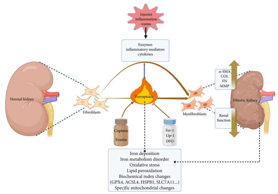 Fig.1 Ferroptosis & renal fibrosis.1
Related Factors of Renal Fibrosis
Renal fibrosis is associated with chronic kidney disease (CKD) and is also the final deleterious outcome of several CKD. Both two affect half of the adults above age 70 and 10 percent of the population in the world. In developed countries, the main inducements of renal fibrosis are type 2 diabetes mellitus and ischemic/hypertensive nephropathy. Additionally, renal fibrosis as a chronic and progressive process influences kidneys during aging and in CKD, regardless of causes. Although no specific therapy yet exists to attenuate fibrosis, a number of current advances have clarified and understood the cellular and molecular mechanisms underlying the disease.
Mechanisms of Renal Fibrosis
Renal fibrosis is an inevitable consequence of the excessive accumulation of ECM that occurs in every type of CKD virtually. This progressive process of fibrosis ultimately brings out the end-stage renal failure, a devastating disorder which requires dialysis or kidney transplantation. Briefly, renal fibrosis represents a failed wound-healing course of kidney tissue after sustained lesions. There are some cellular pathways, including mesangial, fibroblast activation and tubular epithelial-mesenchymal transition (EMT), having been identified as the main approaches for the generation of matrix-producing cells in diseased conditions. Among the various fibrogenic factors, transforming growth factor-beta (TGF-β) is the one who takes a central part to regulate the renal fibrotic process. Though defective matrix degradation might conduce to tissue scarring, the exact action of the matrix-degrading enzymes in the injured kidney has become more complicated. Recent discoveries on endogenous antifibrotic factors have already evolved novel strategies targeted at antagonizing the fibrogenic action of TGF-β/Smad signaling.
Besides TGF-β, platelet-derived growth factor (PDGF) and connective tissue growth factor (CTGF) are confirmed as quintessential players in the fibrosis of most organs. After the initial injury, these pathways synergize with each other to promote fibrosis and activate profibrotic cells. In sum, significant cellular events in tubulointerstitial fibrosis include (1) infiltration of inflammatory cells; (2) fibroblast activation and expansion from various sources; (3) production and deposition of a large amount of ECM components; (4) tubular atrophy and microvascular rarefaction. Convincingly, every one of above mentioned pathologic features can contribute to the relentless progression of fibrosis in its own unique way. Together, they constitute a core set of fibrogenic matters causing the final breaking of renal parenchyma and the loss of kidney function.
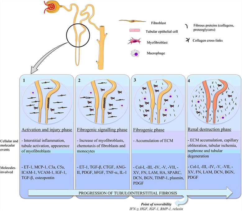 Fig.2 A Visual overview of renal interstitial fibrosis advancement.2
Treatment and IVD Antibody of Renal Fibrosis
Renal fibrosis has been discerned the hallmark of progression of CKD, but, despite intensive research studies, there are currently no beneficial treatments for this condition. Numerous therapeutic interventions perform effective in animal models, however, the transformation of these promising outcomes into humans in the clinical setting still remains a struggling project. There is a hopeful candidate heparanase-1 (HPSE), an endoglycosidase that cleaves heparan sulfate chains and thus takes part in ECM remodeling. As largely described, it acts an essential part in the pathogenesis of cancer and inflammation and participates in the complicated biological progress involved in the onset of diverse renal proteinuric diseases, such as diabetic nephropathy, glomerulonephritis. In addition, HPSE may influence the CKD through its central role in the biological pathway of renal fibrogenesis.
As a leader in the advancing antibody market, Creative Biolabs provides custom IVD antibody development and production services to scientists around the world to support and accelerate their IVD projects. The services we provide include antibody generation, antibody conjugation, and immunoassay development. Our services are fully customized against different targets of different human diseases, including fibrotic kidney diseases.
Our IVD antibody development services target the following biomarkers of renal fibrosis:
References
- Liu, Yi, and Jingyu Wang. "Ferroptosis, a rising force against renal fibrosis." Oxidative medicine and cellular longevity 2022 (2022).
- Genovese, Federica, et al. "The extracellular matrix in the kidney: a source of novel non-invasive biomarkers of kidney fibrosis?" Fibrogenesis & tissue repair 7 (2014): 1-14.
For Research Use Only.