As a leading CRO of antibody generation and development, Creative Biolabs offers application-specific antibody development services to global clients. With experienced scientists and advanced technology, we have established a series of high-quality in vitro diagnostic (IVD) antibody development services against biomarkers of different diseases. Here, we introduce our IVD antibody development services for the CD28 marker.
CD28
CD 28, also known as Tp44, is a T cell surface fraction, and CD28 is a 44 kDa homodimeric glycoprotein linked by a disulfide bond and is a member of the immunoglobulin supergene family. It is present on 95% of human CD4+ T cells, 50% of CD8+ T cells, and most of mouse T cells. The CD28 gene is located on human chromosome 2 and mouse chromosome 1. In recent years, CD28 cDNAs of humans, mice, and rabbits have been successfully cloned and expressed. The human CD28 cDNA encodes an open reading frame containing 220 amino acid residues and has intact membrane protein function. A signal peptide with 18 amino acids at the N-terminus is removed. The mature molecule contains 202 amino acid residues, of which 41 are located in the cytoplasm, 27 were located in the hydrophobic region of the membrane and the rest were located extracellular. There were 5 glycosylation sites in total. Human, murine, and rabbit CD28 exoprotein immunoglobulin heavy chain variable regions have a high degree of homology.
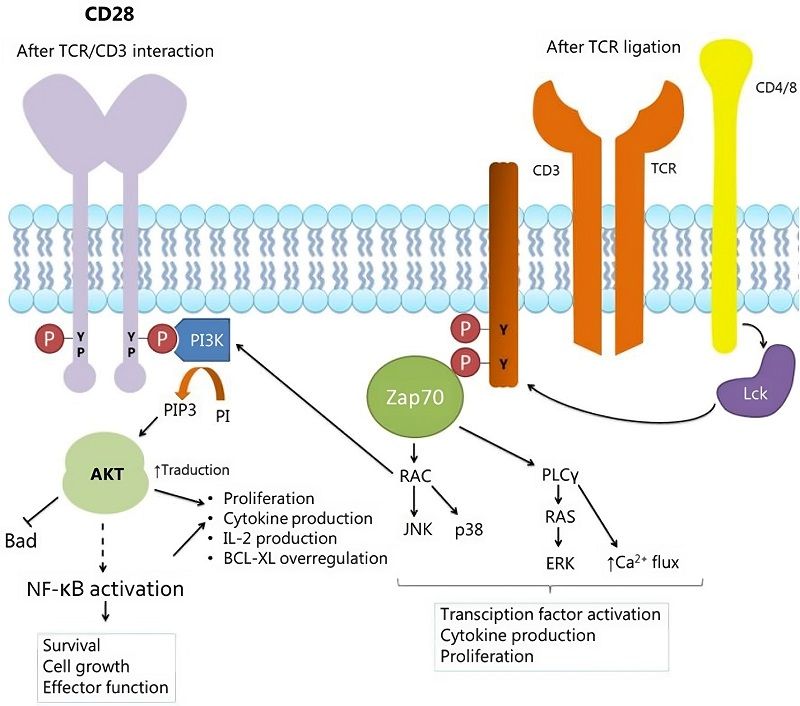 Fig.1 Signaling model of CD28.1
CD28 as Marker of Sepsis
It has been found that the effect of peptidoglycan limiting IL-2 production by T cells can be regulated by the soluble synergistic stimulatory factor B7-2 produced by the T cells themselves, and cytokine production is induced by direct contact between T cells and peptidoglycan-stimulated monocytes. B7-2 specific antibodies prevent the formation of sepsis. In general, B7-2 is expressed within 6 h after the primary stimulation. Studies have found that treatment of CTLA-4 Ig 24 or 48 hours after bacterial invasion will result in a significant reduction in CTLA-4 Ig protection. Treatment of infected animals with a specific CD28 monoclonal antibody (providing a positive co-stimulatory signal resulting in T-cell proliferation and IL-2 secretion) has a higher incidence of sepsis than receiving saline; confirms CD28-B7 co-stimulatory pathway plays a crucial role in the formation of infection-induced abscesses. In addition, antibodies specific to CTLA-4 Ig and CD28 were found to upregulate or downregulate abscess formation, suggesting that T cell activation is a critical step in the host response. Thus, the CD28, CTLA-4/B7 co-stimulatory molecule can be intervened to control bacterial-induced infections.
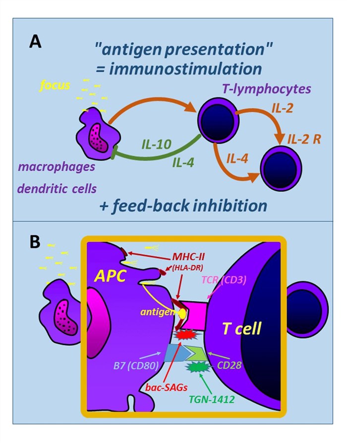 Fig.2 APC-T cell interaction involves CD28. (Gerlach, H., 2016)
IVD Antibody Development Services Targeting CD28 Marker
IVD antibodies have been widely used for disease screening and therapeutic monitoring. As a research partner with years of experience in high-quality antibody development and production, Creative Biolabs offers one-stop solutions from antigen design to antibody pair screening. Besides, we also offer diagnostic immunoassay development services, including feasibility analysis, assay design, assay protocol establishment, assay optimization, and kit production.
Our services can be tailor-designed to adapt to specific project specifications. If you are interested in our services, please do not hesitate to contact us for more details.
References
- Rangel-Sosa, Martha Montserrat, Estuardo Aguilar-Córdova, and Augusto Rojas-Martínez. "Immunotherapy and gene therapy as novel treatments for cancer." Colombia médica 48.3 (2017): 138-147. under Open Access license CC BY 4.0. The image was modified by extracting and using only part of the original image.
- Gerlach, Herwig. "Agents to reduce cytokine storm." F1000Research 5 (2016). under Open Access license CC BY 4.0, without modification.
For Research Use Only.