
Achilles Tendon Injury Modeling & Pharmacodynamics Service
Are you currently facing inconsistent injury replication, high variability in functional recovery data, or challenges in quantifying the regenerative efficacy of your musculoskeletal candidates? Our Achilles Tendon Injury Modeling & Pharmacodynamics Service helps you obtain high-fidelity preclinical data and develop highly effective regenerative therapeutics through standardized surgical protocols and multi-dimensional functional evaluation platforms.
Overview of Achilles Tendon Injury Modeling
The Achilles tendon is the strongest transmitter of mechanical force in the human body, yet its unique physiological environment, characterized by low vascularity and a highly organized Type I collagen matrix, which makes it notoriously difficult to heal. Pathological conditions such as acute ruptures or chronic tendinopathy often result in the formation of disorganized "scar tissue" (Type III collagen) rather than functional regeneration. This leads to persistent mechanical weakness, increased stiffness, and significant clinical morbidity. Creative Biolabs provides the specialized systems needed to analyze these molecular shifts and evaluate the efficacy of next-generation regenerative agents.
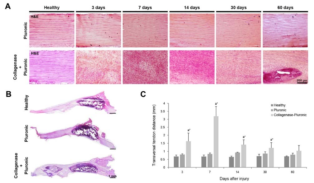 Fig.1 Histological evaluation of rat patellar tendons.1,3
Achilles Tendon Injury Models
Creative Biolabs provides highly reproducible Rat and Rabbit models designed to simulate specific clinical conditions. Our portfolio includes Partial Hemisection Models (ideal for testing radiofrequency or biological adjuncts), Total Transection & Surgical Repair Models (to evaluate post-operative recovery), and Collagenase-Induced Degenerative Models (simulating chronic tendinopathy). By utilizing localized delivery systems like thermo-responsive hydrogels, we ensure the injury remains focal, providing a stable platform for high-precision pharmacodynamic studies and minimizing inter-subject variability.
Evaluation Platform of Our Service
Creative Biolabs offers a suite of analytical capabilities to quantify every stage of the tendon healing process, moving beyond simple histology to provide functional and mechanical validation.
-
Biochemical & Molecular Analysis
- Collagen Density: Hydroxyproline quantification (4-hydroxyproline) via spectrophotometry.
- Gene Expression: qPCR analysis.
- Inflammatory Profiling: Multiplex ELISA.
- Enzymatic Activity: Western Blot for Matrix Metalloproteinases (MMPs).
-
Histopathological Examination
- Fiber Alignment: H&E and Masson's Trichrome staining to evaluate the collagen hierarchy.
- Degeneration Scoring: Bonar and Movin scoring systems for tendinosis assessment.
- Cellular Dynamics: IHC for CD68+ (macrophages) and PCNA+ (proliferating cells).
-
Behavioral & Functional Testing
- Gait Analysis: Automated tracking of stance time, step length, and "Achilles Functional Index" (AFI).
- Nociception: Von Frey filament testing for localized mechanical hypersensitivity.
- Motor Coordination: Rotarod performance and open-field motor assessment.
-
Advanced Imaging & Instrumentation
- High-Resolution Ultrasound: In vivo measurement of Cross-Sectional Area (CSA) and edema levels.
- Ex Vivo Biomechanics: Load-to-failure testing, Ultimate Tensile Strength (UTS), and Stiffness.
- Micro-CT: Precise quantification of intra-tendinous calcifications and bone mineral density.
Key Applications
Our models are designed to simulate critical human indications, including Acute Achilles Ruptures, Chronic Tendinopathy, and Failed Healing (Scarring). We provide the depth required to evaluate a wide range of therapeutic modalities:
- Small Molecules: Anti-inflammatories and targeted protease inhibitors.
- Biologics: Growth factors (GDF-5, BMPs), PRP-based treatments, and therapeutic antibodies.
- Gene Therapies: Viral vectors designed to upregulate tenogenic transcription factors.
- Cell-Based Therapies: Evaluation of MSC or tenocyte integration within the host matrix.
Why Choose Us?
We offer fully validated Rat and Rabbit strains, providing the anatomical flexibility required for both molecular screening and surgical device testing.
Creative Biolabs provides a seamless transition from in vitro tenocyte assays to complex in vivo PD/PK studies and biomechanical validation.
Our PhD-level team ensures rigorous quality management and data integrity, providing the high-resolution evidence needed for regulatory submissions.
Work with Us
- Summarize the project requirements and fill in the information collection form.
- Sign a CDA from both parties to further communicate information, such as targets.
- Select an animal model, discuss experimental design, and determine assay parameters.
- Project costing and project schedule forecasting.
- We provide a detailed project plan, including the required sample quantities, methods, and protocols.
- Both parties confirm the project details and start the project.
- Confirm the timeline of the project.
- We provide periodic results and information on the animal's condition.
- We will work together to make project adjustments as necessary.
- We provide a comprehensive project report promptly.
- We arrange transportation for the produced samples.
- We provide a discussion of the project results and help to arrange the next steps.
- Data storage and archiving.
Frequently Asked Questions
-
Q: Which species is best for testing my biological scaffold?
A: Rabbits are typically preferred for scaffolds or surgical devices due to their larger anatomical size, allowing for more precise surgical integration and biomechanical testing.
-
Q: How do you minimize variability in surgical models?
A: We utilize standardized surgical guides and utilize the contralateral (uninjured) limb as an internal control for every subject.
-
Q: Is longitudinal imaging available?
A: Absolutely. We use high-resolution ultrasound to monitor tendon thickness and edema throughout the study without the need for early euthanasia.
-
Q: How long does a typical regeneration study take?
A: Most studies follow a 7, 14, 28, or 60-day timeline to capture the inflammatory, proliferative, and remodeling phases of repair.
Published Data
Objective: To evaluate the impact of surgical vs. nonsurgical repair on functional torque restoration and muscle preservation.
Model Used: Adult male Rat (Total Achilles Transection).
Results: This model demonstrated high sensitivity in detecting treatment-specific recoveries. Surgical repair yielded a 35% superior increase in plantar flexor torque compared to nonsurgical cohorts. Moreover, the model accurately captured muscle-tendon crosstalk, with surgical intervention halving muscle mass loss (12% vs 21%). The persistence of a post-surgical "functional gap" defines this model as an ideal, rigorous platform for evaluating novel regenerative adjuncts and tissue-engineered constructs in a clinically relevant window.
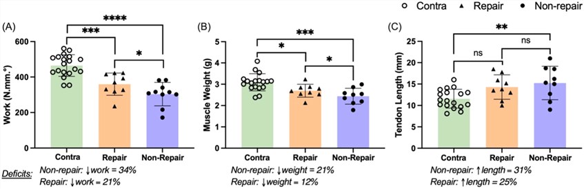 Fig.2 Comparative evaluation of structural and functional outcomes post-treatment of Achilles tendon rupture.2,3
Creative Biolabs is your premier partner for high-precision Achilles Tendon Injury Modeling & Pharmacodynamics. From molecular biomarkers to advanced ex vivo biomechanics, we provide the data your pipeline needs to succeed. Contact our senior scientific team for a detailed technical consultation and project proposal.
References
- Vidal, Laura, et al. "A novel tendon injury model, induced by collagenase administration combined with a thermo-responsive hydrogel in rats, reproduces the pathogenesis of human degenerative tendinopathy." International journal of molecular sciences 25.3 (2024): 1868. DOI: https://doi.org/10.3390/ijms25031868.
- Hammo, Ahmad, et al. "Achilles Tendon Surgical Repair Partially Restores Early Plantar Flexor Structure and Function in a Rat Model." Journal of Orthopaedic Research® 43.4 (2025): 739-745. DOI: https://doi.org/10.1002/jor.26041.
- Distributed under Open Access license CC BY 4.0, without modification.
For Research Use Only.