Epidermophyton floccosum
As a leading company in the field of biological research and drug discovery, Creative Biolabs has gained a wealth of good reputation for successfully completed numerous challenges in antifungal drug discovery. Based on our advanced technology platform and experienced scientists, we are able to provide a series of antifungal drug discovery services against various fungal diseases and related fungi. Here, we describe a pathogenic fungus Epidermophyton floccosum that can cause dermatophytosis.
Introduction
Epidermophyton floccosum (E. floccosum) is an anthropophilic dermatophyte that causes tinea pedis, tinea unguium, tinea corporis, tinea cruris, and tinea manuum in humans. E. floccosum is the only species in the genus Epidermophyton, which belongs to family Arthrodermataceae, order Onygenales, class Eurotiomycetes, subdivision Pezizomycotina, and division Ascomycota. E. floccosum is the third most common cause of tinea pedis worldwide after Trichophyton rubrum and Trichophyton mentagrophytes.
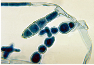 Fig.1 Micrograph of E. floccosum. Distributed under CC BY-SA 4.0, from Wiki, without modification.
Distribution
E. floccosum was originally isolated from a tinea cruris patient in Germany and described as Acrothecium floccosum by Carl Otto Harz in 1870. It distributes throughout the whole world, especially in tropical and subtropical areas. The fungus can survive for years in showers, baths, swimming pools, towels, blankets, sheets, shoes, and other clothing. It is commonly spread by direct contact.
Mycology of E. floccosum
As one of the three dermatophyte fungal genera, the absence of microconidia and the shorter, wide, smooth macroconidia is the distinction of Epidermophyton from the other two genera Microsporum and Trichophyton.
- Macroscopically, colonies are usually slow growing without any specific growth condition in culture. The colonies appear to form a dark yellow to greenish-brown or khaki-colored with a suede-like surface, raised and folded in the center, with a flat periphery and submerged fringe of growth. The opposite color of the colonies is from a burnt orange appearance to sienna brown.
- Microscopically, E. floccosum is a filamentous fungus with septate and hyaline hyphae microscopically. Hyphae are characterized by the smooth, thin-walled, clavate, club-shaped macroconidia and the absence of microconidia, which also is distinguishing feature of this species from the other dermatophytes.
Pathogenesis of E. floccosum
Similar to other fungal dermatophytes, E. floccosum contains keratinase giving it the ability to breakdown keratin within the skin, nails, and hair. A recent case has shown that it can infect eyes causing keratitis. E. floccosum cannot pierce through living tissues of individuals with normal immunity, which results in the infection staying within the nonliving cornified layer (stratum corneum) of host epidermis. Wood's lamp can’t be applied for the diagnosis of the E. floccosum infection as the fungus does not fluoresce under the ultraviolet light.
If you are interested in the fungal disease-related services we investigate, please contact us for more detailed information.
For Research Use Only.