FcγRIIIa Binding Assay
To enhance the understanding of FcγRIIIa binding capabilities for tailored antibody candidates, Creative Biolabs provides an expert service for FcγRIIIa binding testing.
FcγRIIIa Introduction
FcγRIIIa (CD16a), an activating receptor expressed on natural killer cells and mononuclear phagocytes, mediates antibody-dependent cellular cytotoxicity (ADCC) by engaging IgG1 and IgG3 Fc regions. Receptor clustering, driven by multivalent immune complexes, is required for signal transduction due to its intermediate-affinity ligand binding. A functionally significant polymorphism (V176F) generates three allelic forms with differential Fc-binding capacities and clinical responses to therapeutic antibodies. Structural and functional characterization of FcγRIIIa-ligand interactions provides critical insights for optimizing antibody therapeutics, particularly in oncology and autoimmune disorders requiring precise control of cytotoxic activity.
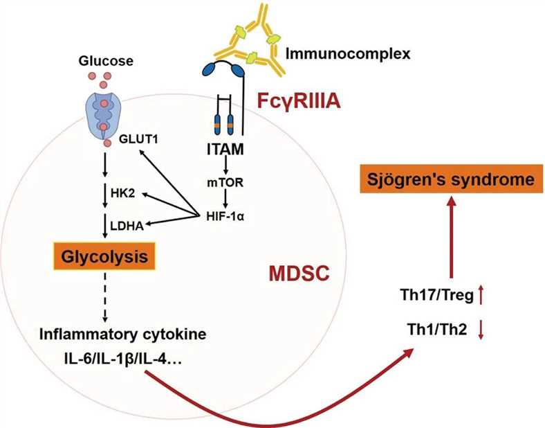
 Fig.1 The signaling pathway of FcγRIIa receptor.1
Our FcγRIIIa Binding Assay
Expanding on the essential insights into FcγRIIIa's function in antibody effectiveness, Creative Biolabs provides an extensive array of methodologies for thorough interaction examination between FcγRIIIa and antibody Fc regions. We offer a comprehensive workflow that starts with customizable assay design aligned with your unique antibody and research goals, and continues with meticulous experimentation using advanced platforms. Anticipate receiving precise and thorough outcomes presented in extensive reports, featuring kinetic parameters, binding affinities, and analyses tailored to specific polymorphisms. Dedicated to fostering a reliable partnership, our knowledgeable scientists offer expert guidance and assistance at every stage, guaranteeing dependable and practical data to enhance your antibody development and optimization initiatives.
Selective Approaches at Creative Biolabs
We offer a series of selective approaches to design for customers' projects. Including but not limited to:
- ELISA
- Flow cytometry
- Surface plasmon resonance / Bio-layer interferometry: real-time, label-free full kinetic analysis.
Service Highlights
- Comprehensive FcγRIIIa coverage
- State-of-the-art technologies
- Customizable assay design
- Reliable and reproducible results
- Comprehensive data reporting
- Fast turnaround times
Case Study
| Objective: | The objective of the investigation is to quantify the binding affinity of sample-1 with FcγRIIIa/CD16a subtypes using the surface plasmon resonance method. | |||||||||||||||||||||||||||||||||
| Assay Format: |
Binding and fitting curves between samples with FcγRIIIa/CD16a (F176): 1:1 binding model; Binding and fitting curves between samples with FcγRIIIa/CD16a (V176): 1:1 binding model; |
|||||||||||||||||||||||||||||||||
| Results: |
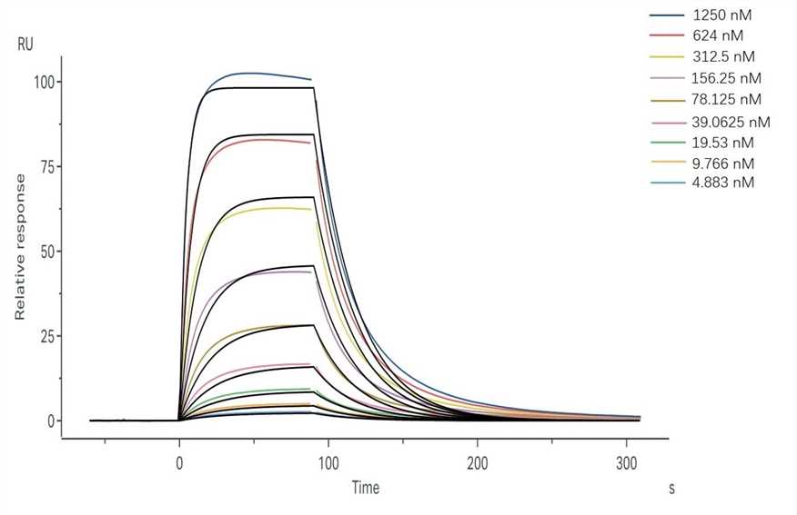
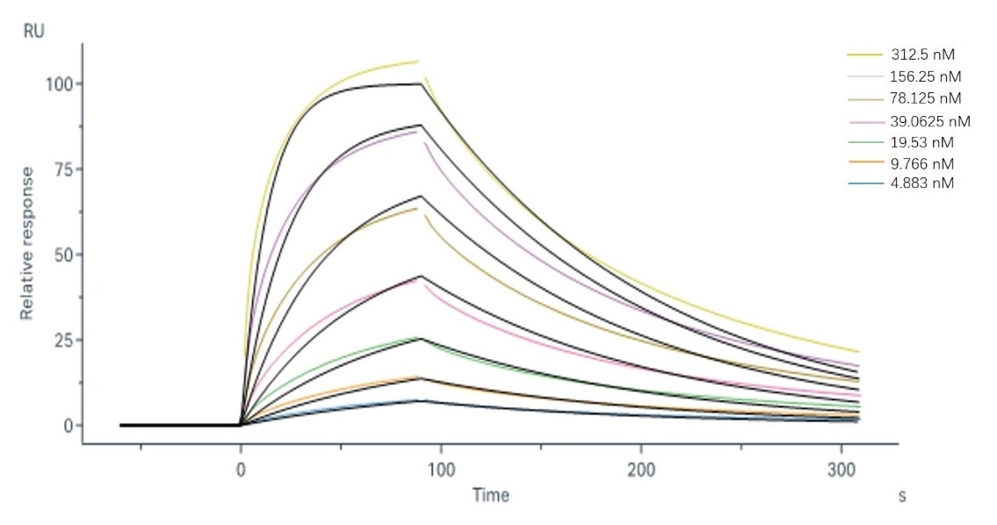
Sample-1 binding to CD16a (F176) The fitting curves for sample-1 binding to CD16a (F176) are shown in Fig.2. Using the 1:1 binding model, the captured CD16a (F176) can bind Sample-1 with an affinity constant of 2.43×10⁻⁷ M. Sample-1 binding to CD16a (V176) The fitting curves for sample-1 binding to CD16a (V176) are shown in Fig.3. Using the 1:1 binding model, the captured CD16a (V176) can bind Sample-1 with an affinity constant of 3.96×10⁻⁸ M. |
|||||||||||||||||||||||||||||||||
|
Table 1. SPR result summary between sample-1 with FcγRIIIa/CD16a subtypes. |
||||||||||||||||||||||||||||||||||
Frequently Asked Questions
Q1: What specimen categories are compatible with your FcγRIIIa binding analysis platform?
A1: Our service accommodates purified immunoglobulins, and Fc-domain fusion constructs, and-following analytical validation-cellular lysates. Project-specific requirements can be addressed through scientific consultation.
Q2: Do your FcγRIIIa interaction assays incorporate soluble or membrane-anchored receptor formats?
A2: Methodologies are available employing both recombinant soluble FcγRIIIa variants and membrane-bound receptor expression systems, accommodating diverse experimental designs.
Related Sections
Our assay platform ensures accurate, sensitive, and reliable measurements, is suitable for automated high-throughput screening, and utilizes clinical-grade positive controls for robust referencing. Serving as a valuable supplement to cell-based ADCC assays, it enables a deeper understanding of lead antibody candidates' mechanisms of action. However, the potential for discrepancies between in vitro results and in vivo efficacy, largely due to interference from endogenous human serum IgGs, necessitates careful interpretation. Furthermore, Creative Biolabs provides Fc engineering services leveraging advanced strategies to enhance ADCC activity by modulating Fc binding characteristics.
If you require further details, do not hesitate to reach out to us or send an inquiry directly.
Reference
- Qi, Jingjing, et al. "FcγRIIIA activation-mediated up-regulation of glycolysis alters MDSCs modulation in CD4+ T cell subsets of Sjögren syndrome." Cell Death & Disease 14.2 (2023): 86. Distributed under Open Access License CC BY 4.0, without modification.
For Research Use Only.

 Fig.2 Binding and fitting curves between sample-1 with FcγRIIIa/CD16a (F176) at different concentrations.
 Fig.3 Binding and fitting curves between sample-1 with FcγRIIIa/CD16a (V176) at different concentrations.