Liver Fibrosis/Cirrhosis Modeling & Pharmacodynamics Services
Creative Biolabs provides a variety of well-established rodent models for evaluating drug efficacy in liver fibrosis/cirrhosis. These models are induced by chemical agents like CCl4, bile duct ligation, or diet induced methods, mimicking the progression from liver injury to advanced fibrosis and cirrhosis. We offer detailed assessments of liver function, histopathology, and molecular biomarkers, enabling comprehensive preclinical testing of potential therapies.
Introduction
Liver fibrosis/cirrhosis is a progressive liver disease characterized by excessive accumulation of extracellular matrix components, leading to scarring and loss of liver function. Fibrosis is the early stage of liver injury, where repeated damage (due to factors like alcohol consumption, viral infections, or non-alcoholic fatty liver disease) triggers an inflammatory response and collagen deposition. If left untreated, it can progress to cirrhosis, which involves extensive fibrosis, liver shrinkage, and impaired function. Cirrhosis can result in complications such as portal hypertension, liver failure, and hepatocellular carcinoma. Common causes of liver fibrosis/cirrhosis include chronic viral hepatitis (Hepatitis B and C), alcohol abuse, non-alcoholic fatty liver disease (NAFLD), and autoimmune liver diseases. Symptoms can include jaundice, ascites, and bleeding disorders.
Disease Models and Applications
Creative Biolabs offers a comprehensive range of well-established rodent models for liver fibrosis/cirrhosis, including models induced by chemical agents like CCl4, bile duct ligation, and diet induced liver injury. These models are carefully designed to replicate the progression of liver fibrosis to cirrhosis, providing an accurate representation of human liver diseases. They are accompanied by thorough evaluations of various parameters such as liver function, histopathological changes, collagen deposition, and serum biomarkers, ensuring the precise assessment of therapeutic candidates during the preclinical phase. Our experienced team of scientists will collaborate with you throughout the project, from experimental design to data interpretation, to ensure high-quality and reliable results. To learn more about the rodent liver fibrosis and cirrhosis models available for preclinical research, please explore the links below:
| Liver Fibrosis/Cirrhosis Models | Simulated Disease | Drug Evaluation Focus | Animal species |
| CCL4 induced Liver Fibrosis/Cirrhosis Model | Liver fibrosis, cirrhosis, liver injury | Anti-fibrotic agents, hepatoprotective drugs, anti-inflammatory drugs | Mouse, Rat, NHPs |
| Thioacetamide (TAA) induced Liver Fibrosis/Cirrhosis Model | Chronic liver injury, cirrhosis, liver fibrosis | Anti-fibrotic drugs, liver regeneration agents, antioxidants | Mouse, Rat |
| Bile Duct Ligation (BDL) induced Primary Biliary Cirrhosis Model | Primary biliary cirrhosis (PBC) | Anti-cholestatic agents, anti-inflammatory drugs, liver regeneration therapies | Mouse, Rat |
| Alpha-Naphthylisothiocyanate (ANIT) induced Primary Biliary Cirrhosis Model | Cholestasis, primary biliary cirrhosis (PBC) | Anti-cholestatic agents, hepatoprotective drugs, anti-inflammatory agents | Mouse, Rat |
| 3,5-Diethoxycarbonyl-1,4-Dihydrocollidine (DDC) induced Primary Biliary Cirrhosis Model | Cholestasis, bile duct injury, primary biliary cirrhosis | Anti-cholestatic drugs, anti-fibrotic agents, hepatoprotective drugs | Mouse |
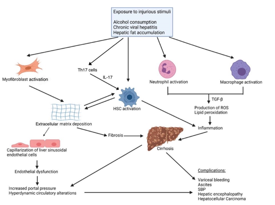 Fig 1. Pathogenesis of fibrosis.1
Measurements
We offer a variety of measurements for evaluating drug efficacy in rodent liver fibrosis/cirrhosis models, utilizing an array of advanced technologies, including but not limited to:
General observations: Body weight, mortality rate, ascites, jaundice, and signs of liver dysfunction.
Histopathology: Liver tissue examination using H&E and Masson's trichrome staining to assess fibrosis severity, liver architecture, and collagen deposition.
Cytokine profiling (e.g., ELISA): Quantification of inflammatory mediators such as TNF-α, IL-6, TGF-β, and IL-1β, which are involved in the progression of liver fibrosis and inflammation.
Hematology analysis and serum biomarkers: Measurement of liver enzymes (ALT, AST), bilirubin levels, and fibrosis markers such as hyaluronic acid and laminin to assess liver function and fibrosis progression.
Gene/protein expression profiling via RT-qPCR and Western blot techniques: Analysis of key fibrosis-related genes (e.g., collagen I, α-SMA, MMPs) and proteins to evaluate the underlying molecular mechanisms of liver fibrosis and cirrhosis.
In addition to the established liver fibrosis and cirrhosis models, our expertise extends to the development of novel animal models tailored to specific research needs, informed by the latest literature and previous studies. Our scientific team is available to assist in experimental design, model selection, and data analysis, ensuring a customized and effective approach to your project at every stage.
Related Services
In addition to liver fibrosis/cirrhosis models, we also offer a wide range of models for other diseases. These models enable comprehensive evaluation across diverse therapeutic areas.
Advantages
- Extensive Expertise and Experience: With years of experience in preclinical research, our team of scientists has deep expertise in liver fibrosis and cirrhosis studies. We are committed to providing accurate, high-quality, and reproducible results to support your research efforts.
- Tailored Solutions for Your Research: We understand that each project is unique. Our models are customizable to fit your specific needs, whether you're focusing on a particular stage of fibrosis, evaluating drug mechanisms, or investigating novel therapeutic approaches.
- Comprehensive and Well-Validated Models: Our rodent models for liver fibrosis and cirrhosis are rigorously validated and designed to replicate key features of human liver diseases. This ensures that your preclinical studies are based on reliable and relevant models, allowing for accurate translation of results.
- State-of-the-Art Technologies: We utilize the latest technologies in histopathology, cytokine profiling, gene expression analysis, and biomarker detection to provide comprehensive insights into disease progression and treatment effects. These advanced techniques enable precise evaluation of therapeutic candidates.
- Expert Support at Every Stage: From experimental design to data interpretation, our experienced team is here to guide you. We provide personalized consultation and assist with model selection, ensuring a smooth and effective research process from start to finish.
- Commitment to Innovation: We continuously invest in the development of new models and techniques, staying at the forefront of scientific advancement to meet the evolving needs of your research.
Work with Us
- Summarize the project requirements and fill in the information collection form.
- Sign a CDA from both parties to further communicate information, such as targets.
- Select an animal model, discuss experimental design, and determine assay parameters.
- Project costing and project schedule forecasting.
- We provide a detailed project plan, including the required sample quantities, methods, and protocols.
- Both parties confirm the project details and start the project.
- Confirm the timeline of the project.
- We provide periodic results and information on the animal's condition.
- We will work together to make project adjustments as necessary.
- We provide a comprehensive project report promptly.
- We arrange transportation for the produced samples.
- We provide a discussion of the project results and help to arrange the next steps.
- Data storage and archiving.
FAQs
-
1. What types of liver fibrosis and cirrhosis models do you offer?
We offer a variety of well-established rodent models for liver fibrosis and cirrhosis, including models induced by chemical agents like CCL4, bile duct ligation, and diet induced liver injury. These models replicate the progression from liver injury to advanced fibrosis and cirrhosis.
-
2. Can your models be customized based on specific research needs?
Yes, our models can be tailored to meet the specific requirements of your research. Whether you are studying early-stage fibrosis or advanced cirrhosis, we can modify the models to best suit your therapeutic objectives.
-
3. What measurements do you provide for liver fibrosis and cirrhosis models?
We provide comprehensive evaluations, including general observations (e.g., body weight, liver function), histopathology (H&E, Masson's trichrome staining), biomarker analysis (liver enzymes, fibrosis markers), and gene/protein expression profiling (collagen, α-SMA, TGF-β).
-
4. What technologies do you use to assess liver injury in your models?
We employ cutting-edge technologies such as immunohistochemistry, ELISA for cytokine profiling, RT-qPCR, and Western blot techniques to assess liver function, inflammation, fibrosis markers, and gene expression related to liver injury.
-
5. How do you ensure the accuracy and reliability of the results?
Our models are rigorously validated to replicate key aspects of human liver diseases, and we follow stringent protocols for data collection and analysis. Additionally, our experienced scientists oversee every stage of your project to ensure reliable and high-quality results.
-
6. How long does it take to get results from your liver fibrosis and cirrhosis models?
The timeline for obtaining results depends on the complexity of the study and the specific model used. Typically, preliminary data is available within a few weeks, with final reports delivered after thorough data analysis.
Published Data
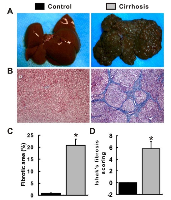 Fig. 2 Establishment of a liver cirrhosis model.2
In this experiment, a liver cirrhosis model was established using thioacetamide (TAA) treatment. Rats in the cirrhosis group displayed noticeable signs of cirrhosis, including reduced body weight, lethargy, and a dull appearance, compared to the control group. All rats administered TAA developed liver cirrhosis without any fatalities related to toxicity. The livers of the control group appeared soft, reddish, and smooth, whereas the livers in the cirrhosis group exhibited a hard texture, blunt edges, brown coloration, and a nodular surface (Fig. 2A). Histological examination using Masson’s trichrome staining further confirmed the presence of liver cirrhosis, showing extensive accumulation of extracellular matrix, disruption of the liver's normal architecture, and formation of pseudolobules (Fig. 2B). The degree of liver fibrosis was quantified using Ishak’s fibrosis scoring, with significantly higher fibrotic areas observed in the cirrhosis group compared to the control group (Fig. 2C). The mean Ishak’s fibrosis score for the cirrhosis group was 5.8, while the control group had a score of 0 (Fig. 2D). These findings confirm the successful establishment of a TAA induced liver cirrhosis model.
References
- Somnay, Kaumudi et al. "Liver Fibrosis Leading to Cirrhosis: Basic Mechanisms and Clinical Perspectives." Biomedicines vol. 12,10 2229. 30 Sep. 2024, DOI:10.3390/biomedicines12102229. Distributed under an Open Access license CC BY 4.0, without modification.
- Lu, Yao-Yao et al. "Cyclooxygenase-2 up-regulates hepatic somatostatin receptor 2 expression." Scientific Reports vol. 8,1 11033. 23 Jul. 2018, doi:10.1038/s41598-018-29349-y. Distributed under an Open Access license CC BY 4.0, without modification.
For Research Use Only.