Non-Alcoholic Steatohepatitis (NASH) Modeling & Pharmacodynamics Services
Creative Biolabs offers a wide range of rodent and non-rodent models for evaluating NASH and related liver diseases. These models allow for comprehensive drug efficacy assessment in areas such as inflammation, lipid metabolism, fibrosis, and liver function. Our expert team provides tailored support in model selection, experimental design, and data analysis, ensuring reliable and high-quality results.
Introduction
Non-Alcoholic Steatohepatitis (NASH) is a progressive liver disease characterized by inflammation, fat accumulation, and liver cell damage in individuals who consume little or no alcohol. It is part of a broader spectrum of non-alcoholic fatty liver disease (NAFLD), which ranges from simple fat accumulation in the liver (steatosis) to more severe forms, including NASH, cirrhosis, and liver failure. NASH is strongly associated with metabolic risk factors such as obesity, type 2 diabetes, dyslipidemia, and insulin resistance. The exact cause of NASH is still not completely understood, but it is thought to result from a combination of factors, including increased fat deposition in the liver, oxidative stress, mitochondrial dysfunction, and an inflammatory response that leads to liver injury. Over time, NASH can progress to fibrosis (scarring of the liver), cirrhosis, and in some cases, liver cancer (hepatocellular carcinoma).
Disease Models and Applications
Creative Biolabs offers a comprehensive range of well-established rodent and non-rodent models for Non-Alcoholic Steatohepatitis (NASH) and related liver disorders. These models are meticulously designed to replicate the pathophysiological features of NASH, including liver fat accumulation, inflammation, oxidative stress, and fibrosis. Our models provide a reliable platform for evaluating therapeutic candidates targeting various aspects of NASH, from early-stage inflammation to advanced liver fibrosis and cirrhosis. Comprehensive evaluations of key parameters, such as liver histopathology, lipid profiles, inflammatory markers, and fibrosis scores, allow for an accurate assessment of drug efficacy during the preclinical phase. Our experienced team of scientists will collaborate closely with you throughout your project, from experimental design to data interpretation, ensuring high-quality, reproducible results. To learn more about the NASH models available for preclinical research, please explore the links below:
| NASH Models | Simulates | Drug Evaluation | Animal species |
| Diet induced Obesity (DIO) Mouse NASH Model | Obesity, Non-Alcoholic Steatohepatitis (NASH), Metabolic Syndrome | Anti-obesity drugs (e.g., orlistat, sibutramine), Lipid-lowering agents (e.g., statins, fibrates), Liver protectants (e.g., pioglitazone) | Mouse |
| High-Fat Diet induced NASH Model | NASH, Fatty Liver Disease, Metabolic Syndrome | Liver protectants (e.g., pioglitazone, statins), Anti-diabetic drugs (e.g., GLP-1 agonists, DPP-4 inhibitors), Anti-obesity drugs | Mouse |
| Methionine Choline-Deficient (MCD) Diet induced NASH Model | NASH, Fatty Liver Disease, Metabolic Dysfunction | Anti-inflammatory drugs, Liver protectants (e.g., pioglitazone, statins), Drugs targeting liver fibrosis and inflammation | Mouse |
| Choline-Deficient L-Amino Acid-Defined (CDAA) Diet induced NASH Model | NASH, Fatty Liver Disease, Liver Fibrosis | Fibrosis inhibitors, Liver protectants, Anti-inflammatory drugs, Antioxidants, Metabolic modulators (e.g., GLP-1 agonists) | Mouse |
| High-Fat & High-Carbohydrate Diet induced NASH Model | NASH, Metabolic Syndrome, Insulin Resistance | Anti-diabetic drugs (e.g., metformin, GLP-1 agonists), Anti-obesity drugs, Liver protectants (e.g., pioglitazone, statins), Lipid-lowering agents | ob/ob, db/db Mouse, Rat, Golden hamster, Rabbit, NHPs |
| High-Fat & High-Cholesterol Diet induced NASH Model | NASH, Metabolic Syndrome, Insulin Resistance | Anti-diabetic drugs (e.g., metformin, GLP-1 agonists), Anti-obesity drugs, Liver protectants (e.g., pioglitazone, statins), Lipid-lowering agents | ob/ob, db/db Mouse, Rat, Golden hamster, Rabbit, NHPs |
| High-Fat & High-Cholesterol Diet & Fructose induced NASH Model | NASH, Fatty Liver Disease, Insulin Resistance | Insulin sensitizers, Anti-diabetic drugs, Liver protectants (e.g., pioglitazone, statins), Anti-inflammatory drugs | Mouse |
| High-Fat & High-Fructose induced NASH Model | NASH, Insulin Resistance, Fatty Liver Disease | Lipid-lowering agents (e.g., statins, omega-3 fatty acids), Anti-diabetic drugs (e.g., GLP-1 agonists, metformin), Anti-inflammatory drugs | Mouse |
| Diethylnitrosamine (DEN) & High-Fat & High-Cholesterol Diet induced NASH Model | NASH, Hepatocellular Carcinoma (HCC), Liver Damage | Anti-cancer drugs, Liver protectants, Anti-inflammatory agents, Fibrosis inhibitors, Lipid-lowering agents | Mouse |
| High-Fat & CCL₄ induced NASH Model | NASH, Liver Fibrosis, Liver Injury | Anti-fibrotic drugs, Anti-inflammatory agents, Liver protectants, Lipid-lowering agents (e.g., statins, fibrates) | Mouse, NHPs |
| Streptozotocin (STZ) & High-Fat induced NASH Model | NASH, Type 2 Diabetes, Hepatic Dysfunction | Anti-diabetic drugs (e.g., metformin, GLP-1 agonists), Liver protectants, Lipid-lowering agents, Insulin sensitizers | Mouse, Rat |
| MC4R KO Mouse Model | Obesity, Metabolic Disorders, NASH | Anti-obesity drugs (e.g., orlistat, sibutramine), GLP-1 agonists, Anti-inflammatory drugs, Lipid-lowering agents (e.g., statins, fibrates) | Mouse |
| LDLR KO Mouse Model | Hyperlipidemia, Atherosclerosis, NASH | Lipid-lowering agents (e.g., statins, omega-3 fatty acids), Anti-inflammatory drugs, Anti-fibrotic drugs | Mouse |
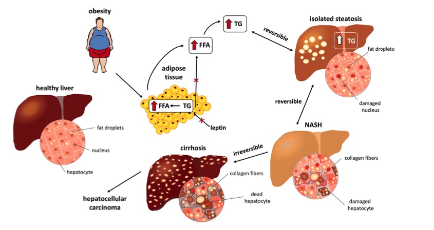 Fig. 1 Stages of NASH.1
Measurements
We offer a variety of measurements for evaluating drug efficacy in non-alcoholic steatohepatitis (NASH) models, utilizing advanced technologies to provide a comprehensive assessment of liver injury, steatosis, inflammation, and fibrosis. These measurements include, but are not limited to:
1. General Observations:
- Body Weight: Monitoring weight changes to assess metabolic effects.
- Mortality Rate: Tracking survival rate during the experiment.
- Food Intake: Changes in food consumption due to drug treatment or disease progression.
- Liver Size and Appearance: Inspection of liver morphology for signs of steatosis and fibrosis.
2. Histological Assessments:
- Hematoxylin and Eosin (H&E) Staining: To assess liver tissue for fat accumulation, inflammation, and fibrosis.
- Oil Red O Staining: For detecting lipid droplets in liver tissues, indicating steatosis.
- Masson's Trichrome Staining: For identifying liver fibrosis and collagen deposition.
- Immunohistochemistry (IHC): Detection of inflammatory markers (e.g., TNF-α, IL-6) and oxidative stress indicators (e.g., 4-HNE).
- TUNEL Assay: To evaluate apoptosis in liver tissue.
3. Cytokine Profiling (e.g., ELISA):
- Inflammatory Mediators: Measurement of cytokines such as TNF-α, IL-6, IL-1β, and chemokines, which are involved in the inflammatory response characteristic of NASH.
- Pro-fibrotic Cytokines: Assessment of TGF-β, PDGF, and MCP-1 for fibrosis-related signaling.
4. Liver Enzyme and Serum Biomarkers:
- Liver Enzymes: Measurement of serum ALT (alanine aminotransferase), AST (aspartate aminotransferase), and ALP (alkaline phosphatase) to assess liver injury and hepatocellular damage.
- Bilirubin Levels: Total and direct bilirubin levels as indicators of liver function.
- Lipid Profile: Serum triglycerides, cholesterol levels, and non-esterified fatty acids (NEFAs) to assess dyslipidemia and fatty liver progression.
5. Gene/Protein Expression Profiling via RT-qPCR and Western Blot Techniques:
- Gene Expression: Quantification of genes involved in fatty acid metabolism (e.g., PPAR-α, SREBP-1c, FAS), inflammation (e.g., TNF-α, IL-6), and fibrosis (e.g., collagen I, TGF-β).
- Protein Expression: Western blot analysis for proteins involved in NASH pathogenesis, such as NF-κB, Nrf2, and fibrotic markers (e.g., α-SMA, collagen).
- Oxidative Stress Markers: Measurement of markers like malondialdehyde (MDA), 8-OHdG, and GPx for evaluating oxidative stress in the liver.
6. Fibrosis Staging:
- Liver Histology: Assessment of fibrosis using semi-quantitative scoring systems (e.g., METAVIR, Ishak scoring system) based on histological features.
- Collagen Content Measurement: Analysis of collagen deposition in liver tissue through histological staining and biochemical assays (e.g., hydroxyproline assay).
7. Ultrasound and Imaging Techniques:
- Liver Ultrasound: To detect liver fat accumulation and fibrosis in vivo.
- Magnetic Resonance Imaging (MRI): For quantitative assessment of liver fat content and fibrosis stages.
Related Services
In addition to non-alcoholic steatohepatitis (NASH) models, we also offer a wide range of models for other diseases. These models enable comprehensive evaluation across diverse therapeutic areas.
Advantages
- Expertise in Disease Models: We specialize in providing comprehensive and well-established rodent models for various diseases, including Non-Alcoholic Steatohepatitis (NASH), metabolic disorders, and other complex pathologies. Our scientific team has extensive experience in designing and customizing models to suit your specific research needs.
- Cutting-Edge Technologies: We utilize advanced diagnostic and measurement technologies, including histology (H&E, Oil Red O, Masson's Trichrome), immunohistochemistry, cytokine profiling, and gene/protein expression profiling (RT-qPCR, Western blot). These technologies ensure a thorough evaluation of disease progression and therapeutic efficacy.
- Customized Approach: Understanding that every project is unique, we offer tailored services that align with your specific research objectives. Whether you are studying early-stage NASH or evaluating advanced fibrosis, our models can be adapted to reflect the conditions you're investigating.
- Comprehensive Biomarker Assessment: Our services include a wide range of biomarkers, including liver enzymes, cytokines, oxidative stress markers, and fibrosis stages. This allows for a holistic evaluation of drug effects, ensuring that you receive the most complete data for decision-making.
- In Vivo Monitoring: We offer advanced in vivo monitoring techniques like ultrasound and MRI to assess liver fat accumulation and fibrosis without the need for repeated invasive procedures, ensuring more reliable and less stressful assessments for the animals.
Work with Us
- Summarize the project requirements and fill in the information collection form.
- Sign a CDA from both parties to further communicate information, such as targets.
- Select an animal model, discuss experimental design, and determine assay parameters.
- Project costing and project schedule forecasting.
- We provide a detailed project plan, including the required sample quantities, methods, and protocols.
- Both parties confirm the project details and start the project.
- Confirm the timeline of the project.
- We provide periodic results and information on the animal's condition.
- We will work together to make project adjustments as necessary.
- We provide a comprehensive project report promptly.
- We arrange transportation for the produced samples.
- We provide a discussion of the project results and help to arrange the next steps.
- Data storage and archiving.
FAQs
-
Q: What types of disease models do you offer?
A: We offer a wide range of rodent and non-rodent disease models, including Non-Alcoholic Steatohepatitis (NASH), obesity, metabolic syndrome, cardiovascular diseases, diabetes, liver fibrosis, and many others. We also provide customized models tailored to specific research needs.
-
Q: What technologies do you use for evaluating drug efficacy?
A: We employ advanced technologies such as histology (H&E, Oil Red O, Masson's Trichrome), immunohistochemistry (IHC), cytokine profiling (ELISA), gene/protein expression profiling (RT-qPCR, Western blot), and in vivo monitoring using ultrasound and MRI. These technologies ensure comprehensive data collection and analysis.
-
Q: Can you customize models to suit specific research needs?
A: Yes, we offer fully customizable animal models. Whether you are studying early or advanced stages of diseases such as NASH or fibrosis, we work with you to design a model that meets your specific research objectives.
-
Q: How do you assess liver function and damage in NASH models?
A: Liver function and damage are assessed using a combination of serum biomarkers (e.g., ALT, AST, liver enzymes), histological analysis, cytokine profiling, and fibrosis staging. These methods help evaluate the extent of liver injury, inflammation, and fibrosis.
-
Q: Do you provide any support for experimental design?
A: Absolutely! Our experienced scientific team is available to assist with experimental design, model selection, and data interpretation. We guide you through every stage of your project to ensure optimal results.
Published Data
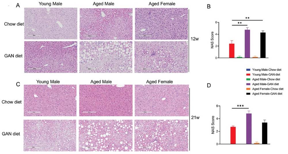 Fig. 2 GAN diet induced Nonalcoholic Steatohepatitis in young and aged mice.2
This article aimed to establish a novel non-alcoholic steatohepatitis (NASH) model using aged mice fed a high-fat, high-sugar, high-cholesterol (GAN) diet. This model was developed to better replicate the rapid progression of human NASH, facilitating preclinical research and drug development. The NAS scoring system for non-alcoholic fatty liver disease (NAFLD) was employed to assess the disease severity in each group of mice. Results showed that aged male mice fed the GAN diet for 12 weeks exhibited a significantly more severe NASH phenotype, characterized by hepatic steatosis, inflammatory cell infiltration, and ballooning degeneration. Their NAS scores were notably higher than those of younger male mice. Aged female mice also displayed exacerbated NASH symptoms after 12 weeks of induction (Fig. 2A and 2B). Additionally, after 21 weeks of GAN diet induction, aged male mice continued to show more severe NASH features (Fig. 2C and 2D).
References
- Kořínková, L et al. "Pathophysiology of NAFLD and NASH in Experimental Models: The Role of Food Intake Regulating Peptides." Frontiers in endocrinology vol. 11 597583. 26 Nov. 2020, DOI:10.3389/fendo.2020.597583. Distributed under an Open Access license CC BY 4.0, without modification.
- Li, Xuecheng et al. "A new NASH model in aged mice with rapid progression of steatohepatitis and fibrosis." PloS one vol. 18,5 e0286257. 25 May. 2023, DOI: 10.1371/journal.pone.0286257. Distributed under an Open Access license CC BY 4.0, without modification.
For Research Use Only.