
Osteoporosis Modeling & Pharmacodynamics Services
Are you currently facing long drug development cycles, high attrition in skeletal drug efficacy, or difficulty in quantifying subtle microarchitectural bone changes? Our Osteoporosis Modeling & Pharmacodynamics Services help you accelerate drug discovery and obtain high-quality, reproducible efficacy data through our validated rodent models and high-resolution Micro-CT analytical platforms. Creative Biolabs streamlines your transition from bench to clinic by providing peer-reviewed, industry-standardized bone mineral density (BMD) depletion frameworks.
Overview of Osteoporosis Modeling
Osteoporosis is a systemic skeletal disorder characterized by low bone mass and microarchitectural deterioration of bone tissue, leading to enhanced bone fragility and a consequent increase in fracture risk. Primarily affecting the aging population and postmenopausal women, it involves an imbalance between osteoclast-mediated bone resorption and osteoblast-mediated bone formation. These conditions often lead to debilitating hip and vertebral fractures. At Creative Biolabs, we provide specialized modeling that accounts for both primary osteoporosis (age-related and postmenopausal) and secondary osteoporosis (drug induced or metabolic), providing a robust foundation for evaluating next-generation anti-resorptives and bone-forming agents.
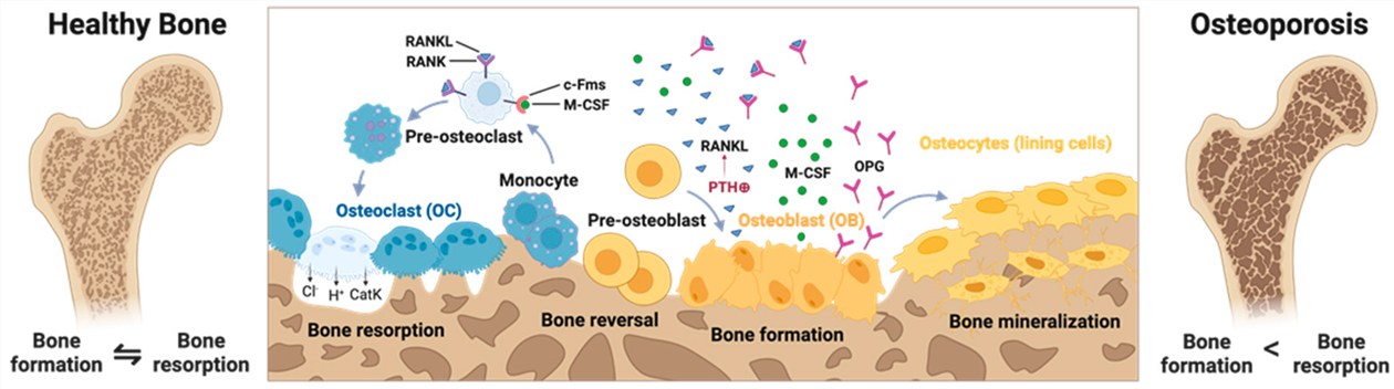 Fig.1 Molecular mechanisms of normal bone and osteoporosis.1
Osteoporosis Disease Models
Creative Biolabs offers a suite of validated models in the Rat and Rabbit species. Our OVX induced model remains the gold standard for postmenopausal research, while our Orchiectomy (ORX) model simulates male-specific bone loss. For secondary osteoporosis, our Glucocorticoid induced (GIOP) and Retinoic Acid induced models provide rapid, predictable bone depletion patterns. These models are characterized by a standardized "loss curve," allowing for precise pharmacological intervention timing.
| Models | Related Disease | Drug Evaluation | Animal Species |
| Orchiectomy induced Osteoporosis Model | Male osteoporosis; Androgen-deficiency induced bone loss; Hypogonadism-related skeletal fragility. | Androgen replacement therapies; Selective Androgen Receptor Modulators (SARMs); Anti-resorptive agents for male patients. | Rat, Rabbit |
| OVX induced Osteoporosis Model | Postmenopausal osteoporosis (Type I); Estrogen-deficiency-mediated bone mass depletion. | Estrogen replacement therapies (ERT); SERMs; Bisphosphonates; RANKL inhibitors; PTH analogs (anabolic agents). | Rat, Rabbit |
| Glucocorticoid induced Osteoporosis Model | Secondary osteoporosis; Steroid induced bone loss (SIBL); Iatrogenic skeletal deterioration from chronic steroid use. | Glucocorticoid-sparing agents; Therapies targeting steroid induced suppression of osteoblast activity; Vitamin D/Calcium analogs. | Rat, Rabbit |
| Retinoic Acid induced Osteoporosis Model | Acute bone loss conditions; Hypervitaminosis A induced skeletal fragility; High-turnover metabolic bone disease. | Rapid screening of novel anti-resorptive candidates; Evaluation of agents targeting accelerated bone resorption pathways. | Rat, Rabbit |
Evaluation Platform of Our Service
Our platform offers deep technical insights into bone quality and cellular metabolism through a standardized parameter list:
-
Biochemical & Molecular Analysis
- Bone Turnover Markers: Serum/Urine assays for CTX-I, NTX-I, PINP, and Osteocalcin.
- Enzymatic Profiling: Alkaline Phosphatase (ALP) and Tartrate-resistant Acid Phosphatase (TRAP) activity.
- Molecular Signaling: Western Blot and qPCR for RANKL/OPG ratio and Wnt/β-catenin pathway analysis.
-
Histopathological Examination
- Standard Staining: H&E and Masson's Trichrome for tissue integrity.
- Cellular Scoring: TRAP staining for osteoclast counting and IHC for protein localization.
- Dynamic Histomorphometry: Calcein double-labeling to measure bone formation rates.
-
Behavioral & Functional Testing
- Biomechanical Strength: Three-point bending and femoral neck compression tests.
- Locomotor Assessment: Gait analysis and grip strength to evaluate functional recovery.
-
Advanced Imaging & Instrumentation
- High-Res Micro-CT: 3D analysis of BV/TV, Tb.N, Tb.Th, and Tb.Sp.
- DXA Scanning: In vivo longitudinal Bone Mineral Density (BMD) measurement.
Key Applications
Our models are designed to simulate a wide range of clinical indications and support the evaluation of diverse therapeutic modalities:
- Indications: Postmenopausal Osteoporosis, Senile (Age-Related) Osteoporosis, Steroid induced Bone Loss, and Fracture Healing.
- Therapeutic Agents: Small molecule inhibitors, monoclonal antibodies (biologics), gene therapies targeting skeletal pathways, and cell-based regenerative therapies.
Why Choose Us?
We utilize a wide range of validated Rat and Rabbit strains, specifically selected for their human-analogous bone remodeling cycles.
We provide a seamless transition from initial in vitro screening to comprehensive in vivo PD/PK studies, ensuring data consistency across your development cycle.
Our PhD-level team maintains rigorous quality management systems and provides data packages that are audit-ready for regulatory submissions.
Work with Us
- Summarize the project requirements and fill in the information collection form.
- Sign a CDA from both parties to further communicate information, such as targets.
- Select an animal model, discuss experimental design, and determine assay parameters.
- Project costing and project schedule forecasting.
- We provide a detailed project plan, including the required sample quantities, methods, and protocols.
- Both parties confirm the project details and start the project.
- Confirm the timeline of the project.
- We provide periodic results and information on the animal's condition.
- We will work together to make project adjustments as necessary.
- We provide a comprehensive project report promptly.
- We arrange transportation for the produced samples.
- We provide a discussion of the project results and help to arrange the next steps.
- Data storage and archiving.
Frequently Asked Questions
-
Q: Can you perform longitudinal tracking without euthanizing animals?
A: Yes, Creative Biolabs utilizes in vivo DXA and Micro-CT scanning, allowing us to track BMD changes in the same animal over time, which enhances statistical power.
-
Q: What is the typical turnaround time for a standard efficacy study?
A: Depending on the model, most studies range from 8 to 12 weeks. We provide interim reports at key milestones to keep your project on track.
-
Q: Do you offer biomechanical testing to prove fracture risk reduction?
A: Yes, we perform rigorous ex vivo three-point bending and compression tests to provide the "gold standard" proof of bone strength.
-
Q: Are your protocols compliant with international regulatory filings?
A: Yes, our studies are conducted under strict quality controls designed to meet the documentation requirements of major global regulatory bodies.
Published Data
Objective: To validate the temporal progression of bone mineral density depletion in the Wistar model to identify the optimal "intervention window."
Model Used: Ovariectomized (OVX) Wistar Rat (Female, 12 weeks old).
Results: This model exhibits exceptional longitudinal predictability, with a high-fidelity transition from osteopenia to clinical osteoporosis between days 30 and 40 post-surgery. Linear degradation of Tb.N and BV/TV provides a sensitive, quantifiable baseline for pharmacological assessment. Notably, the identification of Day 30 as the optimal "pivot point" allows for an accelerated and cost-effective timeline for evaluating preventative therapies. These attributes establish the OVX Wistar model as a superior, high-throughput platform for capturing bone-protective kinetics with high clinical translational value.
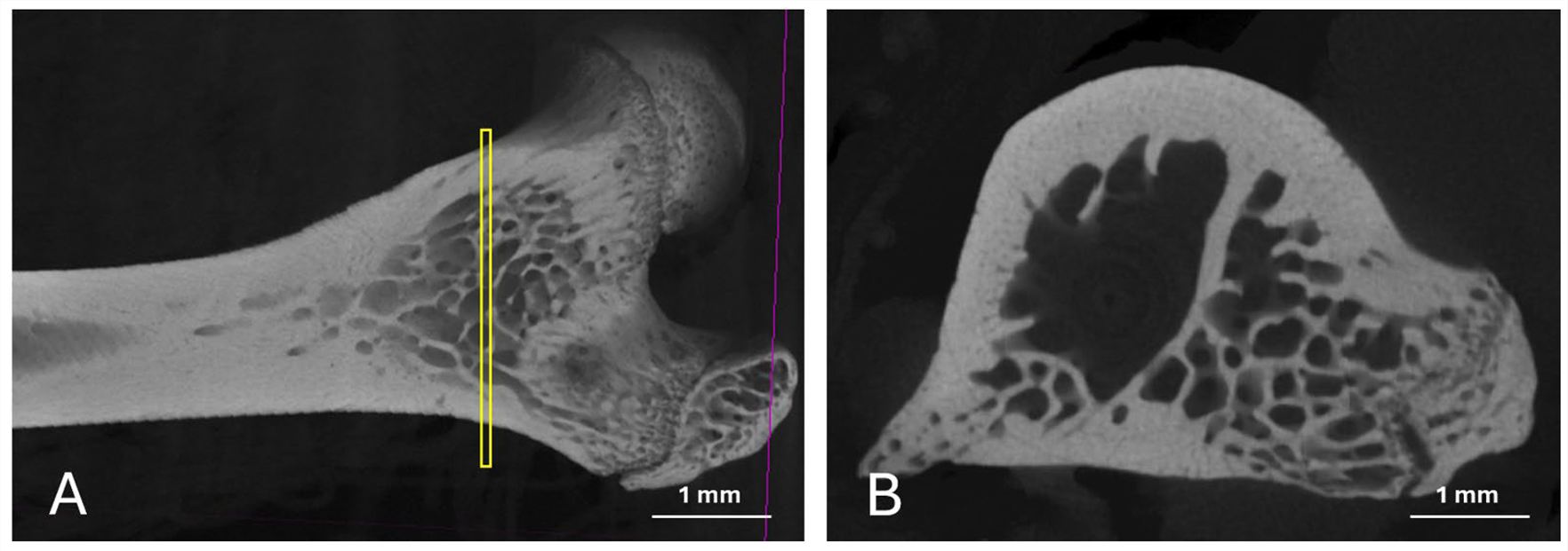 Fig.2 Typical images depicting the sagittal cut position for trabeculae counting and density assessment.2
Creative Biolabs is dedicated to providing the technical precision and scientific narrative required to move your skeletal therapies forward. Our validated Rat and Rabbit models, combined with high-resolution imaging and biochemical expertise, offer a robust foundation for your next breakthrough. Our specialists are standing by to provide a tailored optimization roadmap for your study.
References
- Plank, Johannes, et al. "Modelling Osteoporosis in Pre-Clinical Research—Challenges, Trends and New Approaches." Cells 14.21 (2025): 1649. Distributed under Open Access license CC BY 4.0. The image has been modified; only part of the original image. DOI: https://doi.org/10.3390/cells14211649.
- Pedroso, Artur Lage, et al. "The Validation of an Experimental Model in Wistar Female Rats to Study Osteopenia and Osteoporosis." Bioengineering 12.7 (2025): 702. Distributed under Open Access license CC BY 4.0, without modification. DOI: https://doi.org/10.3390/bioengineering12070702.
For Research Use Only.