Ovariectomy (OVX) induced Osteoporosis in Rats
Osteoporosis is a disease characterized by low bone mass and structural deterioration of bone tissue, leading to bone fragility and an increased susceptibility to fractures. The incidence of osteoporosis increases with age and occurs most frequently in postmenopausal women due to the dramatic estrogen withdrawal associated with menopause. Animal models of postmenopausal bone loss induced with ovariectomy provided by Creative Biolabs present characteristics of bone loss and its sequelae resembles those found in postmenopausal women in some respects due to ovarian hormone deficiency. As a consequence, this model is widely applied for studying problems that are relevant to postmenopausal bone loss.
The Ovariectomized Rat Model
For induction of bone loss, female rats with 3 months of age are ovariectomized. Although the skeletal growth is continued, studies have proved that the rats are sexually mature at this time of age and is sufficient to induce bone loss. About 3 months after ovariectomy, robust ovariectomy-induced pathological changes to the bone structure at different sites, including the proximal tibia, femur neck, and lumbar vertebrae.
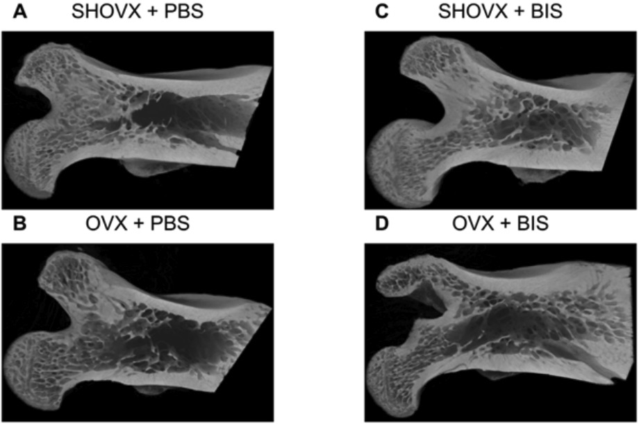 Fig. 1 Characterization of the ovariectomy induced osteoporotic phenotype in rats. 1
Similarities Shared with Postmenopausal Bone Loss
- The ovariectomy-induced bone loss in the rat share many similarities with women postmenopausal bone loss.
- Firstly, there is a reduction in skeletal mass caused by an imbalance between bone resorption and bone formation, with the former exceeding the latter.
- Moreover, there is a rapid phase of bone loss in the beginning, and then the rate tends to slow down.
- Thirdly, the bone loss tends to be greater to cancellous than cortical bone.
- In measuring the calcium content, usually, the intestinal absorption of calcium is declined. When treated with therapies such as estrogen, similar skeletal responses can be observed.
Features of the Ovariectomized Rat Model
- It is a preclinical experimental model approved by the Food and Drug Administration (FDA).
- It is extensively used for not only studying how the decreased production of endogenous estrogen by the ovaries leads to postmenopausal osteoporosis but how potential therapeutic agents can prevent bone loss in this state.
Assessments
In testing the therapeutic efficacy of osteoporosis drug candidates, we are capable of conducting all kinds of assessments as well as providing assistance in data interpretation. Our battery of assessments including but not limited to:
- Bone turnover markers
- Serum/Urine calcium
- Serum phosphorus
- Bone mineral density (BMD)
- Trabecular number, thickness
- Trabecular bone volume fraction
- Micro-CT
- Histopathological analysis
Additionally, other examples of rodent musculoskeletal disease models that you may be interested include:
- Monosodium Iodoacetate (MIA)-Induced Osteoarthritis Model
- Surgically-Induced Osteoarthritis (OA) Models
- Fracture Models
- Bone Defect Models
- Orchiectomy-Induced Osteoporosis Model
- Adjuvant-Induced Arthritis (AIA) Rodent Model
- Collagen-Induced Arthritis (CIA) Rodent Model
- Collagen Antibody-Induced Arthritis (CAIA) Model
Supported by a team of experts with years of experience in the field of musculoskeletal research and surgeons who are skilled in animal surgery, Creative Biolabs is proud to provide a comprehensive list of drug efficacy testing services. Particularly, we are willing to share our expertise and work with you to design and develop experimental models with the highest clinical relevance. If you're interested in our services, contact us for more information.
Reference
- Lotz, Ethan M., et al. "Ibandronate treatment before and after implant insertion impairs osseointegration in aged rats with ovariectomy induced osteoporosis." Journal of Bone and Mineral Research Plus 3.7 (2019): e10184. Distributed under Open Access license CC BY 4.0. The image was modified by extracting and using A-D part of the original image.
For Research Use Only.