Rodent Photothrombotic Ischemia Model
Creative Biolabs has established a number of well-characterized rodent stroke models of permanent, transient, focal and global ischemia, mechanically or chemically induced, to aid in the development of new neuroprotective strategies. The photothrombotic stroke model as presented here is a model of focal ischemia that consistently induces a stroke injury of the desired size in any cortical or subcortical site in a very reproducible and minimally invasive manner.
Induction of Photothrombotic Ischemia Model
The photothrombotic stroke model was initially proposed by Rosenblum and El-Sabban in 1977 and was later improved by Watson in 1985 in rat brain, laying the basis of the current model. This model induces an ischemic damage within a given cortical area by means of photo-activation of a previously injected light-sensitive dye. Following illumination, the dye is activated and produces singlet oxygen that damages components of endothelial cell membranes, with subsequent platelet aggregation and thrombi formation, which eventually determines the interruption of local blood flow.
Briefly, a photoactive dye (e.g., Rose Bengal, erythrosine B) is injected intraperitoneally (mice) or intravenously (rats) and enters the bloodstream. Then, the intact skull is irradiated within minutes by a light beam at a specific wavelength, which activates the dye and generates oxygen radicals that lead to endothelial damage, platelet activation, and aggregation in both pial and intraparenchymal vessels within the irradiated area. The evaluation of ischemic damage can be quickly accomplished by triphenyl-tetrazolium chloride (TTC) or cresyl violet staining.
Features of Photothrombotic Ischemia Model
- This model produces infarction of small size and well-delimited boundaries, which is highly advantageous for precise cell characterization or functional studies.
- This model is further characterized by rapid progression of ischemic cell death.
- The light source can be applied to the intact skull with no need of craniotomy, which allows targeting of any cortical area of interest in a reproducible and non-invasive way.
- It requires minimal surgical intervention and the lesion is highly reproducible.
- This model shows a high survival rate.
- This approach is not time-consuming, few expensive and can be quickly learned as it is not technically demanding as opposed to other models, including the filament model.
- It is particularly suitable for studying cellular and molecular responses underlying brain plasticity in transgenic mice.
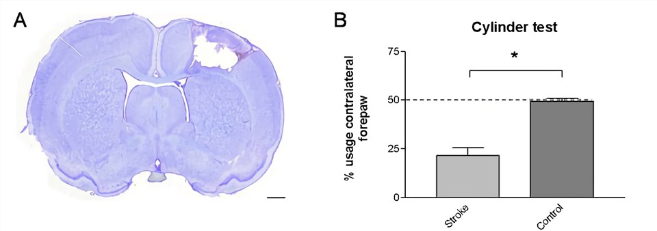 Fig.1 Photothrombotic stroke model. (A) Cresyl violet staining of the photothrombotic stroke region. (B) Cylinder test. (Vandeputte et al. 2010)1, 2
Assessments
For the study of potential neuroprotective and therapeutic agents in human stroke, our neurological disease research platform offers an array of behavioral tests, behavioral recuperation tests, and histological assessments, including but not limited to:
- Behavioral tests (e.g., motor function, cognition, social behavior)
- Cerebral blood-flow and vessels imaging (transcranial laser Doppler)
- Infarct volume measurement (magnetic resonance imaging (MRI), tetrazolium chloride (TTC))
- Histology
Creative Biolabs also offers other stroke & brain ischemia rodent models that you may be interested in:
- Middle Cerebral Artery Occlusion (MCAO) Model (transient/permanent)
- Endothelin-1 Model
- Global Ischemic Four-Vessel Occlusion (4-VO) Rat Model
Creative Biolabs is recognized for the excellent quality of its Non-Clinical studies and the expertise of our scientific team ensures the highest quality data for our partners. In addition to stroke & brain ischemia studies, we can also provide specialized efficacy models in other neurological system diseases. The comprehensive list of rodent neurological models is placed below for your review:
If you do not see the model you are interested in, just contact us and Creative Biolabs can customize an existing model or work with you to develop a new model with the highest clinical relevance.
References
- Vandeputte, C.; et al. Automated quantitative gait analysis in animal models of movement disorders[J]. BMC Neuroscience. 2010, 11(1):92.
- under Open Access license CC BY 4.0, without modification.
For Research Use Only.