
Tibial Fracture Modeling & Pharmacodynamics Service
Are you currently facing high failure rates in fracture union studies, inconsistent trauma modeling, or difficulties in correlating biological healing with functional recovery? Our Tibial Fracture Modeling & Pharmacodynamics Service helps you obtain highly reproducible longitudinal data and validate therapeutic efficacy through precision-engineered Rat models and multi-dimensional analysis. By utilizing advanced three-point bending and high-energy impact technologies, Creative Biolabs ensures your drug candidates are tested in the most clinically relevant environments to streamline your path to the clinic.
Overview of Tibial Fracture Modeling
The skeletal system is a dynamic physiological structure governed by a delicate balance of osteoblast-driven formation and osteoclast-mediated resorption. Tibial fractures, particularly those involving high-energy trauma or open wounds, pose a significant clinical challenge due to the risk of "nonunion", the permanent failure of bone fusion. These conditions are often exacerbated by vascular disruption, chronic inflammation, and soft tissue damage. Understanding the interaction between mechanical stability and the biological secretome (growth factors like VEGF and TGF-β) is critical for developing next-generation therapies for delayed union, osteoporosis-related fractures, and traumatic bone loss.
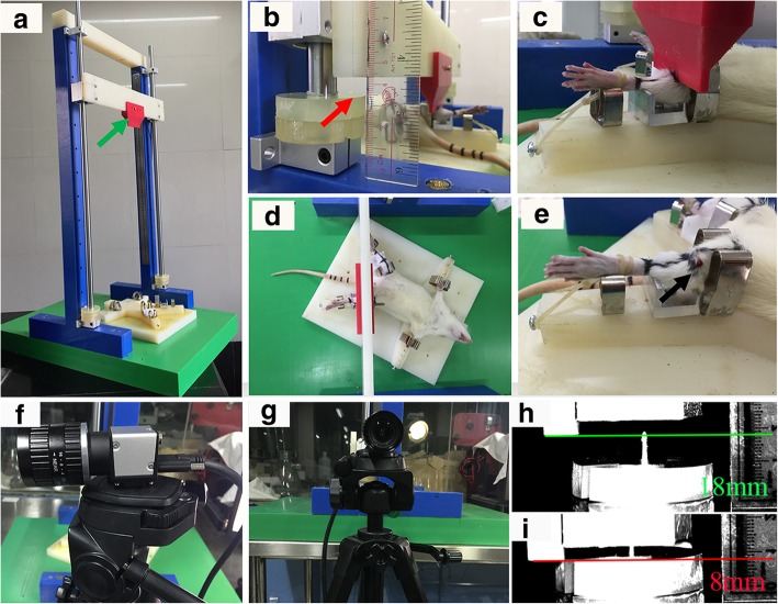 Fig.1 The device and procedure for a standard moderate open fracture (Type II).1,3
Tibial Fracture Models
Creative Biolabs offers a suite of validated Rat models designed to simulate diverse clinical scenarios. These include the Optimized Closed Fracture Model using precision three-point bending for secondary healing studies, the High-Energy Open Fracture Model to evaluate biologically compromised sites, and the Tibial Osteotomy with Plate Fixation Model for longitudinal functional recovery tracking. Our models ensure minimal unintended comminution and high reproducibility in fracture gap width.
Evaluation Platform of Our Service
Creative Biolabs provides a multi-dimensional analysis platform to quantify every stage of the bone healing cascade. Our technical parameters include:
-
Biochemical & Molecular Analysis:
- Systemic & Local Cytokine Assays (TNF-α, IL-1β, IL-10, TGF-β).
- Protein Expression via Western Blot (Osteocalcin, RUNX2).
- Gene Expression via qPCR for osteogenic markers.
- Serum Biomarker Monitoring (α-2-Macroglobulin).
-
Histopathological Examination:
- Standard Staining (H&E, Masson's Trichrome) for collagen and cell infiltration.
- Immunohistochemistry (IHC) for angiogenesis (CD31) and apoptosis (Caspase-3).
- Semi-quantitative Scoring (Emery Fracture Healing System).
-
Behavioral & Functional Testing:
- Longitudinal Gait Analysis (Duty factor, temporal symmetry, swing speed).
- Static Weight Bearing (Incapacitance testing).
- Mechanical Testing (Torsional and four-point bending strength).
-
Advanced Imaging & Instrumentation:
- High-Resolution Micro-CT (Bone Volume/Total Volume, Trabecular thickness).
- Longitudinal Radiography (Callus area and cortical bridging scores).
- In vivo Bone Mineral Density (BMD) measurement.
Key Applications
Our models simulate a wide range of human indications, including Traumatic Bone Nonunion, Delayed Union, Osteoporotic Fractures, and Post-Surgical Recovery. Creative Biolabs supports the evaluation of various therapeutic modalities:
- Small Molecules: Systemic or local osteogenic regulators.
- Biologics: Growth factor concentrates, monoclonal antibodies, and cytokines.
- Gene & Cell Therapies: Viral vectors for bone repair and mesenchymal stem cell (MSC) applications.
- Innovative Delivery Systems: Nanoparticles, hydrogels, and bio-scaffolds.
Why Choose Us?
We utilize highly validated Rat strains, ensuring translational relevance and robust historical control data.
Creative Biolabs provides a seamless transition from initial in vitro biocompatibility to in vivo pharmacodynamics and PK/PD modeling, all under one roof.
Our PhD-led team brings over 20 years of experience in orthopedic biology, backed by rigorous quality management systems and a commitment to data integrity.
Work with Us
- Summarize the project requirements and fill in the information collection form.
- Sign a CDA from both parties to further communicate information, such as targets.
- Select an animal model, discuss experimental design, and determine assay parameters.
- Project costing and project schedule forecasting.
- We provide a detailed project plan, including the required sample quantities, methods, and protocols.
- Both parties confirm the project details and start the project.
- Confirm the timeline of the project.
- We provide periodic results and information on the animal's condition.
- We will work together to make project adjustments as necessary.
- We provide a comprehensive project report promptly.
- We arrange transportation for the produced samples.
- We provide a discussion of the project results and help to arrange the next steps.
- Data storage and archiving.
Frequently Asked Questions
-
Q: How does Creative Biolabs ensure the reproducibility of the fracture gap in Rat models?
A: We utilize modified precision pliers or standardized guillotine apparatuses with adjustable buffer discs to control the energy transfer, ensuring a consistent fracture type every time.
-
Q: Can your platform evaluate both biological fusion and functional recovery?
A: Yes. We integrate longitudinal micro-CT with advanced gait analysis to ensure that therapeutic effects are measured by both structural bridging and the animal's return to normal weight-bearing.
-
Q: Is it possible to test local delivery systems like hydrogels or scaffolds?
A: Absolutely. Our surgical team is an expert in performing intramedullary fixation or plate fixation, allowing for the precise implantation of local therapeutic carriers directly at the fracture site.
-
Q: What is the typical turnaround time for a longitudinal study?
A: Studies typically range from 4 to 12 weeks, depending on whether you are measuring initial callus formation or long-term remodeling and functional restoration.
Published Data
Objective: To correlate biological bone healing with functional gait recovery following internal fixation of a tibial osteotomy.
Model Used: Rat Tibial Osteotomy with Plate Fixation and longitudinal monitoring.
Results: This high-fidelity model demonstrated that micro-CT-confirmed cortical bridging and BMD increases correlate precisely with kinetic recovery. Specifically, longitudinal gait analysis showed that "hindlimb duty factor imbalance" and "temporal symmetry" act as sensitive, non-invasive indicators of the healing stages. By bridging the gap between structural integrity and functional performance, this platform serves as a validated, real-time pharmacodynamic tool to accelerate drug efficacy screening in orthopedic and regenerative medicine.
Creative Biolabs is your premier partner for Tibial Fracture Modeling & Pharmacodynamics. Whether you are developing a novel small molecule or a complex cell therapy, our platform provides the precision and depth required to advance your research with confidence. Contact our specialist team today to receive a customized study design and quote.
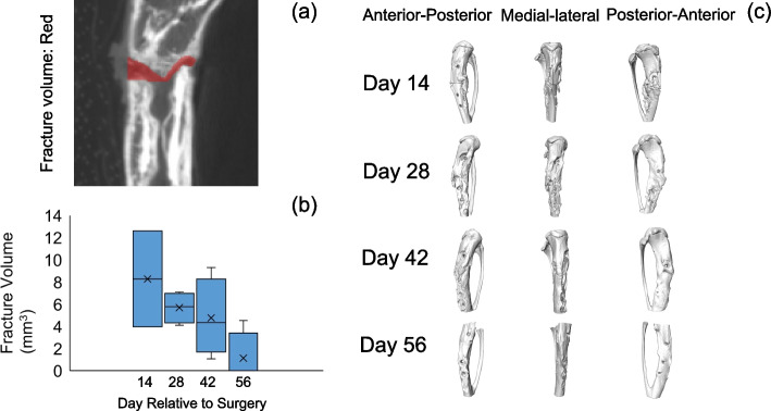 Fig.2 Quantitative assessment of tibia fracture volume at the osteotomy site2,3
References
- Shi, Enxian, et al. "A novel rat model of tibial fracture for trauma researches: a combination of different types of fractures and soft tissue injuries." Journal of Orthopaedic Surgery and Research 14.1 (2019): 333. Distributed under Open Access license CC BY 4.0, without modification. DOI: https://doi.org/10.1186/s13018-019-1386-4.
- Fan, Yingfang, et al. "A longitudinal rat model for assessing postoperative recovery and bone healing following tibial osteotomy and plate fixation." BMC Musculoskeletal Disorders 24.1 (2023): 854. DOI: https://doi.org/10.1186/s12891-023-06942-5.
- Distributed under Open Access license CC BY 4.0, without modification.
For Research Use Only.