Trichophyton rubrum
As a leading company in the field of biological research and drug discovery, Creative Biolabs has gained a wealth of good reputation for successfully completed numerous challenges in antifungal drug discovery. Based on our advanced technology platform and experienced scientists, we are able to provide a series of antifungal drug discovery services against various fungal diseases and related fungi. Here, we describe a pathogenic fungus Trichophyton rubrum that can cause dermatophytosis.
Introduction
Trichophyton rubrum (T. rubrum) is the most common agent of dermatomycoses isolated from humans. It belongs to genus Trichophyton, family Arthrodermataceae, order Onygenales, class Eurotiomycetes, subphylum Pezizomycotina, and phylum Ascomycota. T. rubrum was first described as Epidermophyton rubrum by Castellani in 1910. It has been considered as a cause of chronic tinea corporis in the late nineteenth century. Since then, T. rubrum has spread throughout the world as the etiological agent of tinea unguium and tinea pedis.
Distribution
Out of all the dermatophyte infections, the species T. rubrum is the most prevalent as it is endemic throughout the world except for Antarctica. It is an exclusively clonal, anthropophilic saprotroph that inhabits the upper layers of dead skin, which primarily causes dermatophytosis as tinea pedis, tinea unguium, tinea corporis, and tinea capitis worldwide.
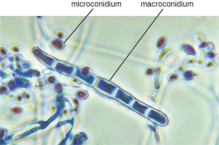 Fig.1 Micrograph of T. rubrum. Distributed under CC BY-SA 4.0, from Wiki, without modification.
Dimorphism of T. rubrum
Two types of T. rubrum commonly described are T. rubrum downy type and T. rubrum granular type.
- The downy type is formed by slender, clavate or club-shaped microconidia and does not make macroconidia under the microscope.
- The granular type makes a large number of clavate (oblong with thick ends) to pyriform (pear-shaped) microconidia and scanty to moderate macroconidia. Macroconidia are usually absent, but once present, they are smooth, thin-walled multiseptate, slender and cylindrical to bacillus-shaped.
Colonies of T. rubrum are white and cottony on the surface and have a reverse side that ranges from yellow-brown to wine-red. Most cultures have been identified to be granular strains, which include numerous microconidia and small spores produced from asexual reproduction.
Pathology of T. rubrum
T. rubrum is a keratinophilic filamentous fungus. During infection, T. rubrum can produce and secrete proteolytic enzymes as important virulence factors. It is indicated that these proteases have the ability to digest the keratins in tissues such as nail and skin stratum corneum into short peptides and amino acids for the assimilation by T. rubrum and facilitate the invasion into keratinized tissues. Since T. rubrum is highly sensitive to environmental pH conditions, the secretion of the proteases depend on the pH content of keratins and is optimal in acidic conditions. Moreover, more than half of the T. rubrum genome sequence is composed of proteases, mostly keratinases. The T. rubrum genome has now been organized into five chromosomes, in which 43 unique nuclear-encoded genes have been analyzed and approximately more than half are proteases.
If you are interested in the fungal disease-related services we investigate, please contact us for more detailed information.
For Research Use Only.