AAV Vector Design for Achromatopsia
Achromatic color ablation (achromatopsia) belongs to complete cone dysfunction, which is exactly the opposite of night blindness (rod cell dysfunction). Patients are especially dark, photophobic, and manifested as blindness seriously, with only the distinction between light and dark, and no color difference. Moreover, there are symptoms such as poor vision, amblyopia, central dark spots, and oscillating nystagmus for achromatopsia patients. It is the most serious type of color vision disorder and is less common in patients. Pathogenic genes are mainly CNGA3, CNGB3, GNAT2, PDE6H, PDE6C and ATF6. Adeno-associated virus (AAV) vectors in gene CNGA3 delivery therapy for achromatopsia have entered phase I clinical trials. Based on the latest research progress, Creative Biolabs provides comprehensive gene therapy technologies and services to improve the symptoms of achromatopsia.
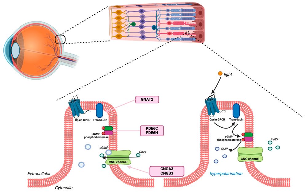 Figure 1. Achromatopsia genes encoding proteins in the phototransduction cascade.
AAV Vectors for Gene Therapy of Achromatopsia
Currently, scientists have designed AAV vectors by carrying cone-specific promoter fragments to offset the congenital cone photoreceptor dysfunction. So far, similar studies have been registered for four applications. Encouragingly, three of them are already in clinical trials. Besides, scientists have used AAV vectors in eight animal models to genetically treat the disease in preclinical research.
AAV Vectors Restore Retinal Function in the Sheep Model
Achromatopsia is mainly caused by mutations in the CNGA3 gene. According to reports, inserting a complete CNGA3 gene from humans into the AAV vector, following successfully delivered into the sheep subretinal can get a long-term recovery of visual function. The sheep in the drug-administered group used a short time to pass through the maze and rarely collided, compared with the control group.
AAV Vectors Improve Vision in the Mouse Model
The pathogenic genes are GNAT1, OPN4 and CNGA3 in the mouse model for achromatopsia. Researchers have demonstrated that the transplant of AAV vectors to increase CNGA3 gene expression can effectively increase the function of the retina. Electroretinogram (ERG) and optomotor response (OMR) are clinically used to evaluate retinal function. After AAV vector is injected into the mouse retina, ERG rescue was about 40 to 50 percent. Moreover, ERG rescue was up to 80% after three weeks of continuous administration, which means that gene therapy using AAV vectors can improve the condition of achromatopsia in the mouse model.
Our Achromatopsia Gene Therapy Development Services
At Creative Biolabs, we provide comprehensive and customizable solutions to support the development of gene therapies for Achromatopsia (ACHM). Our integrated platform is specifically tailored to address the unique biological and translational challenges associated with cone photoreceptor restoration.
1. Target Gene Selection & Optimization
- CNGA3 / CNGB3 construct design
- Codon optimization for cone expression
- Promoter selection (cone-specific promoters)
- Expression cassette engineering
2. AAV Vector Engineering
Efficient delivery of therapeutic genes to cone photoreceptors is critical for successful treatment. Our AAV engineering capabilities focus on optimizing retinal tropism, transduction efficiency, and long-term expression.
| Service Component | Description | Key Benefits |
|---|---|---|
| Serotype Screening | Evaluation of AAV serotypes (e.g., AAV2, AAV5, AAV8, AAV9, and engineered variants). | Identifies the most efficient vector for cone targeting. |
| Capsid Engineering | Development of modified or novel capsids to enhance retinal penetration and reduce immunogenicity. | Improves delivery efficiency and safety. |
| Vector Genome Design | Construction of single-stranded or self-complementary AAV genomes. | Enables rapid and robust gene expression. |
| Tropism Validation | Assessment of vector specificity in retinal explants and animal models. | Ensures accurate targeting of cone photoreceptors. |
3. AAV Capsid Selection and Engineering
Efficient transduction of cone photoreceptors depends heavily on the choice and optimization of the AAV capsid. Our platform evaluates both naturally occurring serotypes and engineered variants to identify the most suitable vector for retinal delivery.
| Capsid | Key Characteristics | Advantages | Typical Use |
|---|---|---|---|
| AAV2 | Well-characterized retinal vector | Extensive validation | Subretinal delivery |
| AAV5 | Enhanced photoreceptor tropism | Efficient cone transduction | ACHM programs |
| AAV8 | High transduction efficiency | Rapid onset of expression | Preclinical studies |
| AAV9 | Broad tissue tropism | Potential for alternative delivery routes | Exploratory applications |
| Engineered Capsids | e.g., AAV2-7m8, Anc80L65 | Improved penetration and reduced immunogenicity | Intravitreal or next-generation therapies |
Our capsid engineering services include directed evolution, rational design, and immune evasion strategies, ensuring optimal vector performance tailored to specific therapeutic goals.
4. Vector Production and Quality Control
To ensure clinical readiness, we offer scalable vector production and rigorous quality assessment in compliance with regulatory standards.
| Service | Description | Outcome |
|---|---|---|
| Research-Grade Production | Small-scale vector generation for preclinical studies. | Rapid evaluation of candidate constructs. |
| Manufacturing | Production of high-titer AAV vectors using scalable and reproducible methods. | Supports research applications. |
| Purification Technologies | Iodixanol gradient and chromatography-based purification. | High purity and potency. |
| Quality Control Testing | Includes identity, purity, potency, sterility, and endotoxin testing. | Ensures regulatory compliance. |
| Stability Studies | Assessment under various storage conditions. | Determines shelf life and handling requirements. |
Comprehensive AAV Vector Design Strategies for ACHM
At Creative Biolabs, vector design is a multi-dimensional process. We meticulously engineer every component of the AAV cassette to maximize therapeutic index.
01. Gene Cargo and Sequence Optimization
The foundation of a successful AAV vector is the therapeutic transgene. Our molecular biology team optimizes the genetic payload for maximal stability and expression:
- Codon Optimization
- Intron and UTR Engineering
- Dual AAV Vector Systems
02. Tissue-Specific Promoter Selection and Engineering
Ubiquitous promoters (like CMV or CAG) can lead to ectopic expression in retinal pigment epithelium (RPE) or rod cells, potentially causing toxicity or metabolic stress. We focus on cone-specific promoters:
- hGRK1 (Human Rhodopsin Kinase 1) Promoter
- PR1.7 Promoter
- M-Opsin and S-Opsin Promoters
Why Partner with Creative Biolabs for AAV Design?
Developing an ophthalmic gene therapy is a complex, high-stakes endeavor. Creative Biolabs serves as your dedicated wet-lab extension, offering critical advantages:
- Deep Ophthalmic Expertise: We possess specialized knowledge in retinal anatomy, photoreceptor biology, and the unique challenges of ocular gene delivery.
- End-to-End Customization: We do not rely on a "one-size-fits-all" approach. Every promoter, capsid, and regulatory element is selected specifically for your ACHM target and intended strategy.
- Data Integrity and IP Security: All research is conducted under strict confidentiality (CDA/NDA). The custom vector designs, optimized sequences, and resulting preclinical data belong entirely to you.
- Accelerated Timelines: By integrating bioinformatics design, cloning, virus production, and in vivo testing under one roof, we eliminate the delays associated with managing multiple vendors.
What You Receive
Partnering with Creative Biolabs provides you with a comprehensive and regulatory-ready package tailored to support every stage of Achromatopsia gene therapy development. Our deliverables are designed to accelerate your program from concept to clinical translation with clarity and efficiency.
Optimized Vector Constructs
You will receive codon-optimized AAV constructs targeting key Achromatopsia genes such as CNGA3 and CNGB3. Each design incorporates cone-specific promoters and regulatory elements to ensure precise and durable expression in photoreceptors.
High-Quality AAV Vector Production
We provide high-titer, research-grade AAV vectors suitable for both in vitro and in vivo studies. For programs advancing toward the clinic, we also offer guidance for manufacturing and scalable production strategies. Formulation and storage recommendations are supplied to maintain vector stability.
Comprehensive Analytical and Quality Data
Each vector is accompanied by rigorous quality control documentation, including assessments of identity, purity, genome integrity, potency, and sterility. Additional characterization such as transduction efficiency and endotoxin levels ensures reliability and regulatory compliance.
In Vitro and In Vivo Validation Support
Our deliverables include efficacy and safety data generated from relevant retinal models. In vitro studies confirm transgene expression and functional rescue, while in vivo evaluations provide insights into visual function, biodistribution, and ocular safety, supporting translational advancement.
Customer Reviews

Frequently Asked Questions (FAQ)
Q: What is the optimal AAV serotype for achromatopsia in humans?
A: Based on current data, AAV2.7m8 and AAV2.A834 have shown the highest foveal cone transduction in NHPs. However, the optimal serotype may depend on your specific transgene and promoter. We offer a capsid screening service where we test up to 10 variants in your preferred model (mouse, dog, or NHP retinal explants) and provide a ranked recommendation.
Q: How do you address pre-existing neutralizing antibodies to AAV?
A: We offer two strategies: (1) seroprevalence screening of patient populations to select a low-prevalence serotype; (2) engineered capsid variants (e.g., AAV2.7YF, AAV2.NN) that evade neutralizing antibodies. We also provide empty capsid competition assays to predict clinical immunogenicity.
Q: Which genes are most commonly targeted in AAV-based therapies for Achromatopsia?
A: The majority of Achromatopsia cases are caused by mutations in the CNGA3 and CNGB3 genes, making them the primary targets for AAV-mediated gene replacement. Other less common targets include GNAT2, PDE6C, PDE6H, and ATF6. Our team provides customized vector design tailored to the specific genetic mutation and therapeutic objectives of each project.
Q: What is the most effective AAV serotype for delivering genes to cone photoreceptors?
A: Several AAV serotypes have demonstrated efficient transduction of cone photoreceptors, including AAV2, AAV5, and AAV8, with AAV2 and AAV5 being the most widely used in clinical studies. Additionally, engineered capsids such as Anc80L65 or AAV2-7m8 can further enhance retinal penetration and transduction efficiency. We perform serotype screening to identify the optimal vector for each application.
Contact Us
AAV vectors are widely used in the treatment of ophthalmic diseases due to its non-toxic advantages. Creative Biolabs provides a full range of vector design and gene delivery services, especially in eye diseases, for example, achromatopsia. Please contact us in time for more information and we are happy to offer the best service and guidance for you.
Reference
- Michalakis S, Schön C, Becirovic E, et al. Gene therapy for achromatopsia. The journal of gene medicine, 2017, 19(3): e2944. https://doi.org/10.3390/ijms25179739 Distributed under Open Access license CC BY 4.0, without modification.