In Situ Hybridization Detection Service for Circular RNA (circRNA)
Introduction
RNA research faces challenges like long cycles, complex data analysis, and precise non-coding RNA localization. Our In Situ Hybridization Detection for Circular RNAs service accelerates research with advanced ISH methodologies, providing high-resolution spatial data. It visualizes and quantifies circRNA expression in cells/tissues, offers spatial context missing in bulk sequencing, aiding validation, function study, and biomarker identification.
Discover How We Can Help - Request a Consultation
Custom In Situ Hybridization Techniques for Circular RNAs
Circular RNAs (circRNAs) represent a newly recognized category of non-coding RNA molecules, distinguished by their distinctive, covalently closed-loop structure. This feature grants them exceptional stability and resistance to exonuclease digestion, making them attractive as stable biomarkers for a wide range of diseases. While high-throughput sequencing has enabled the discovery of thousands of circRNAs, it is In Situ Hybridization (ISH) that provides the crucial spatial context, allowing researchers to visualize where these molecules are expressed within a cell or tissue.
Fluorescence ISH (FISH)
Using probes labeled with fluorescent molecules (e.g., FITC, Cy3, Cy5), it visualizes signals via fluorescence or confocal microscopy, a common choice for circRNA detection. It enables simultaneous detection of multiple circRNAs or co-localization with other molecules (e.g., mRNA, proteins) with high resolution, clarifying subcellular circRNA localization. Probes are optimized as gapmers, multi-probe sets, or LNA-modified (for better hybridization and lower background). It applies to intracellular localization, co-expression studies, and detecting circRNA distribution differences in pathological tissues.
Chromogenic ISH (CISH)
It converts probe labels into colored precipitates (e.g., brown, purple) via enzymatic reactions (e.g., HRP, AP) for observation under a standard light microscope. Signals are stable (results are storable long-term), and they require no fluorescence microscope, making them suitable for routine labs or batch testing of clinical samples. It is ideal for qualitative/semi-quantitative analysis of circRNAs in clinical pathological samples (e.g., circRNA expression differences in tumor tissues), facilitating correlation with pathological morphology.
CircFISH (Circular Fluorescent In Situ Hybridization)
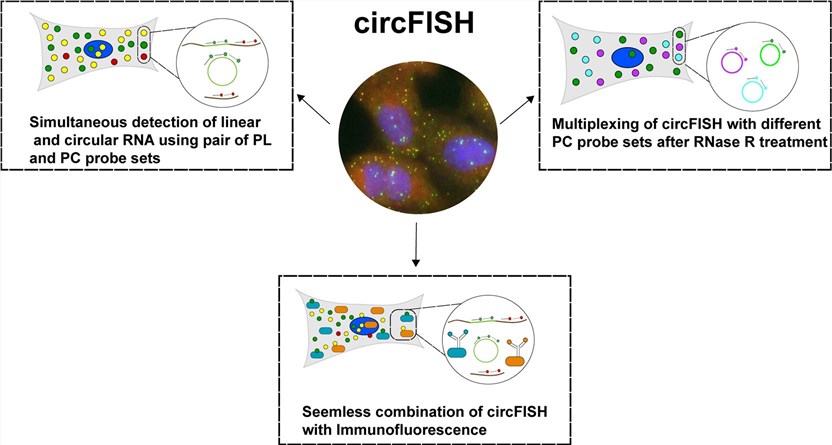 Fig.1 CricFISH can simultaneously detect both linear and circular RNAs and can also be combined with multiple immunological detection methods.1
It is a new technique improved from single-molecule FISH (smFISH), enabling simultaneous imaging of linear RNA and circular RNA (circRNA) in fixed cells and tissues with single-molecule resolution. Its core principle involves designing two sets of specific probes: PC probes (Probe Circular) targeting circRNA-specific sequences, and PL probes (Probe Linear) targeting sequences unique to linear RNA. With different fluorescent labels, linear RNA shows mixed-color signals under fluorescence microscopy, while circRNA shows single fluorescent signals (binding only PC probes), achieving their distinction.
Workflow
Required Starting Materials:
- The specific circRNA sequence of interest, including the back-splice junction (BSJ) sequence.
- The type of sample you will provide, such as fresh frozen tissue sections, formalin-fixed paraffin-embedded (FFPE) blocks, or cell pellets.
- A clear description of your project objectives, including any specific cell types or disease states you wish to investigate.
- Key Steps Involved
-
Probe Design and Synthesis
Leveraging advanced bioinformatic tools, we design a custom oligonucleotide probe cocktail. It is typically 15-25 nt in length and has LNA modifications to enhance binding stability, specifically targeting the unique back-splice junction of your circRNA. To minimize cross-reactivity, we perform rigorous in silico validation against linear mRNA homologs and optimize probe GC content to avoid non-specific binding. -
Sample Preparation and Optimization
Your provided tissues or cells undergo tailored preparation. They are fixed with 4% paraformaldehyde or antigen-retrieved, then permeabilized with 0.1% Triton X-100 to ensure probe access to intracellular circRNAs. Pre-hybridization steps further block non-specific binding sites, with adjustments for sample type. -
Hybridization and Signal Amplification
Samples are hybridized with fluorescently labeled probes under optimized conditions. The process is conducted at 42°C for 16-20 hours in a humidified chamber to balance specificity and binding efficiency. For low-abundance circRNAs, we employ a branched DNA signal amplification system. Secondary probes bind to primary probes, triggering cascade signal multiplication. We avoid enzyme-based systems to eliminate background noise from endogenous enzymes. -
Imaging and Data Analysis
High-resolution images are captured using a confocal microscope to ensure single-molecule resolution. Quantitative analysis includes counting individual circRNA molecules per cell, measuring signal intensity, and determining subcellular localization via co-staining with a nuclear marker or cytoplasmic marker. Localization percentages are calculated. -
Final Report Generation
Our comprehensive report includes detailed methods, raw/processed data, high-resolution merged images, and expert interpretation. It highlights key findings and contextualizes results with functional implications. -
Final Deliverables: Upon project completion, you will receive:
- A comprehensive project report with detailed methodology and data analysis.
- High-resolution, publication-ready images of your stained samples.
- Quantitative data tables on circRNA expression levels and localization.
- Estimated Timeframe: The usual timeframe for this service is 2-4 weeks, varying based on the project complexity, sample type, and the number of targets under analysis.
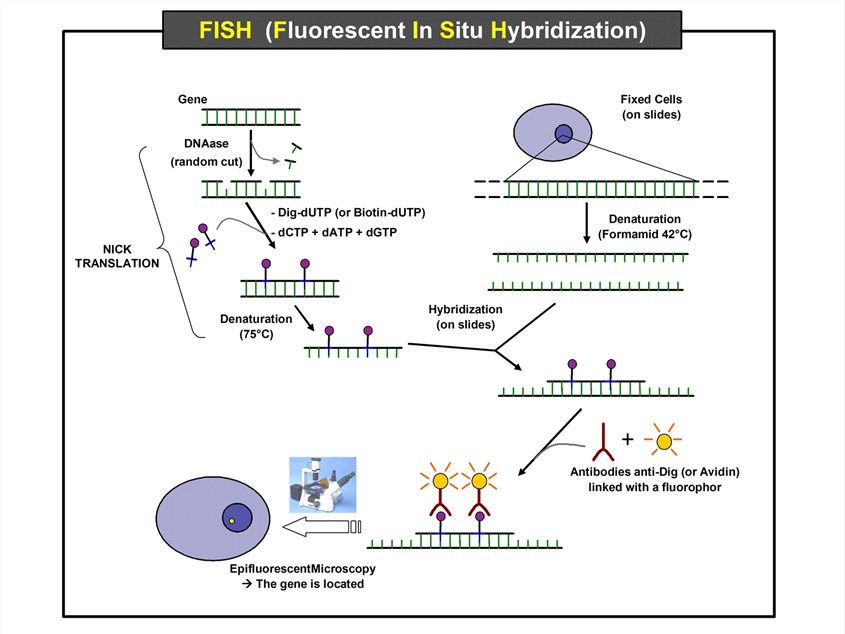 Fig.2 Schematic diagram of the FISH experiment for locating genes in the cell nucleus.Distributed under CC BY-SA 3.0, from Wiki, without modification.
Click to get more details on the workflow
What we can offer
At Creative Biolabs, our In Situ Hybridization Detection for Circular RNAs service meets your precise research needs. With specialized expertise, we offer scientifically sound, highly customizable solutions. We partner with you from consultation to data delivery to ensure project success.
Expertly Designed Probes
We offer customized probe design targeting the unique back-splice junction (BSJ) of your circRNA, ensuring unmatched specificity and sensitivity to distinguish it from its linear mRNA counterpart.
Customization for Any Sample Type
Whether your project involves fresh frozen tissue, archival formalin-fixed paraffin-embedded (FFPE) sections, or cultured cell lines, our protocols are optimized to deliver high-quality results.
Comprehensive Data Delivery
We provide more than just images; our deliverables include quantitative data on expression levels, co-localization analysis, and professional interpretation from our team of scientific experts.
Scalable Multiplexing Solutions
Our platform allows for the simultaneous detection of multiple circRNAs within the same sample, enabling complex co-localization studies and a deeper understanding of molecular interactions.
Seamless Workflow and Support
We provide end-to-end support, from initial project discussion and experimental design to final data interpretation, ensuring a smooth and efficient process that saves you valuable time and resources.
Experience the Creative Biolabs Advantage - Get a Quote Today
Case Study
| circFISH | |
|---|---|
|
|
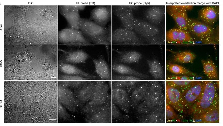
| Fig.3 Schematic diagram of circFISH imaging both linear and circular target RNAs simultaneously.1 | |
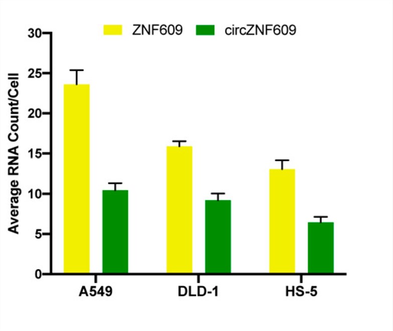
| Target RNA Quantification | Localization of target RNA |
|
|
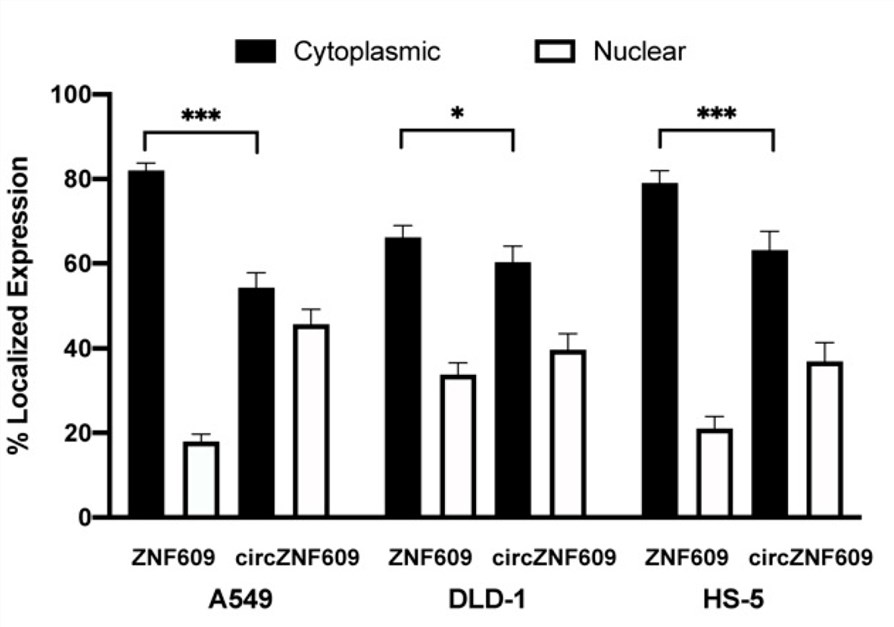
| Fig.4 The linear and circular target RNA signals were quantified after analysis by MATLAB.1 | Fig.5 Average relative nuclear and cytoplasmic localization of linear and circular target RNAs in different cell lines.1 |
Customer Reviews

FAQs
How do you differentiate between circular RNA and its linear counterpart?
We specifically design probes that target the back-splice junction (BSJ), the unique sequence formed by the head-to-tail splicing event that creates the circRNA. This ensures high specificity and prevents cross-detection of the linear mRNA.
Can your service be used for both cell lines and tissue samples?
Yes, our protocol is fully optimized for a wide variety of sample types, including cultured cells, fresh frozen tissues, and formalin-fixed paraffin-embedded (FFPE) blocks. We will work with you to ensure your samples are handled appropriately for the best results.
Is your service compatible with multiplexing for detecting multiple circRNAs at once?
Our advanced platform and probe design allow for multiplexing, enabling the simultaneous detection of multiple circRNAs within the same sample. This provides valuable insights into co-localization and potential interactions.
Creative Biolabs' In Situ Hybridization Detection for Circular RNAs service offers a powerful solution for advancing your research. From custom probe design to high-resolution data analysis, we provide a seamless, professional experience tailored to your project.
Contact Our Team for More Information and to Discuss Your Project
Reference
- Koppula, Aakash, et al. "CircFISH: a novel method for the simultaneous imaging of linear and circular RNAs." Cancers 14.2 (2022): 428. https://doi.org/10.3390/cancers14020428. Distributed under Open Access license CC BY 4.0, some figures were cropped.