Determination Service of Nucleic Acid Concentration
At Creative Biolabs, we've meticulously designed a suite of advanced services that integrate micro-scale spectrophotometry, highly specific fluorescence analysis, and automated capillary electrophoresis to eliminate the uncertainties prevalent in critical experiments. We provide actionable, high-fidelity metrics, such as specific double-stranded DNA concentrations, comprehensive purity ratios, and objective integrity scores, enabling researchers to confidently conduct next-generation sequencing, quantitative amplification, and gene editing projects.
What is Nucleic Acid Quantification?
Nucleic acid quantification refers to measuring the concentration and purity of DNA or RNA in a sample, a crucial step in molecular biology experiments. Common methods include ultraviolet-visible spectrophotometry, which estimates concentration by measuring the absorbance of the sample at a wavelength of 260 nm; and fluorescence-based detection methods, which typically use fluorescent dyes that bind to nucleic acids, thus offering higher sensitivity and specificity. Other techniques include real-time quantitative PCR (qPCR), which is highly sensitive and provides information on the sample's amplification capacity.
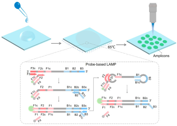 Figure 1. The diagram of probe-based LAMP reaction on a paper membrane for nucleic acid quantification.1
DNA Quantification
DNA quantification aims to determine the absolute abundance and purity of double-stranded DNA, single-stranded DNA, or genomic DNA, which is crucial for genome sequencing, quantitative amplification, and clone construction validation.
- For genomic DNA (gDNA), integrity is a critical quality indicator because highly sheared gDNA leads to uneven sequencing coverage. For NGS libraries, accurate fragment size distribution determines sequencing efficiency.
- Automated Fragment Analysis: Used to accurately determine fragment size, molar concentration, and homogeneity in libraries or sheared DNA samples, ensuring compliance with sequencer requirements.
RNA Quantification
RNA quantification, especially total RNA or messenger RNA (mRNA), is more complex than DNA quantification because RNA is highly degraded, and its integrity has a profound impact on its function.
- For RNA sequencing and other transcriptomics applications, RNA integrity is far more critical than its concentration. Degraded RNA leads to 3' end amplification bias and inaccurate gene expression data.
- ssRNA Specific Quantification: Since RNA is typically present in single-stranded form in samples, we use fluorescent dyes specifically designed for single-stranded nucleic acids to avoid false high readings caused by DNA or other contaminants, thus providing more accurate ssRNA concentrations.
Applications of Nucleic Acid Concentration Determination
Accurate NA quantification underpins all major biological disciplines, acting as a critical point of standardization:
| Downstream Application | Input Requirement | Impact of Inaccurate Quantification |
|---|---|---|
| Next-Generation Sequencing (NGS) | Highly precise ng-to-µg quantities, high purity. | Library preparation failure; skewed library representation; loss of rare variants; non-GEO compliant data. |
| Quantitative PCR (qPCR/RT-qPCR) | Consistent, known input mass for comparative analysis. | Inaccurate absolute or relative gene expression values ($C_t$ value shift); inability to normalize data correctly. |
| Microarray Analysis | Known and consistent starting concentration for labeling efficiency. | Uneven labeling; high background; false differential expression calls. |
| Digital PCR (dPCR) | Known concentration close to the platform's optimum loading concentration. | Sub-optimal partitioning; coalescence leading to underestimation of absolute copy number. |
| Cloning and Transfection | Accurate molar ratios for ligation; consistent DNA amounts for cell uptake. | Low ligation efficiency; cytotoxicity or low transfection rates. |
Nucleic Acid Concentrations Techniques
Molecular biology laboratories rely on three different and complementary techniques to determine nucleic acid concentrations, each with its own mechanistic advantages and limitations.
A. Ultraviolet Spectrophotometry
- Mechanism: This method utilizes the Beer-Lambert law, which states that the absorbance at a specific wavelength is directly proportional to the concentration of the absorbing substance. Nucleic acids, due to their nitrogenous base structure, absorb light most strongly at a wavelength of 260 nm.
- Main Limitation: Ultraviolet spectrophotometry lacks specificity. Any contaminants that absorb light at or near 260 nm (e.g., nucleotides, phenols, guanidine groups) will cause the calculated concentration to be overestimated, leading to an overestimation of the actual functional nucleic acid content.
B. Fluorescence-Based Quantification Methods
- Principle: Fluorescence methods offer significantly higher specificity and sensitivity. They use specific fluorescent dyes that bind to nucleic acids; more importantly, these dyes typically bind selectively to target molecules (e.g., double-stranded DNA or RNA).
- Platform: Qubit fluorometers are standard laboratory equipment, while microplate readers can be used for high-throughput, customized fluorescence detection.
- High Sensitivity: Limit of Quantitation (LOQ) is typically in the picogram range, making it suitable for ultra-low concentration samples.
C. Digital PCR (dPCR)
- Mechanism: dPCR is the gold standard for absolute nucleic acid quantification. Samples are split into thousands of microreactions (droplets or microwells) and then subjected to endpoint PCR.
- Advantages: Digital PCR is inherently absolute and requires no standard curve. It exhibits high tolerance to inhibitors and provides the most accurate measurement of actual amplifiable copy number, crucial for determining functional concentrations.
Benefits and Limitations of Concentrations Technologies
The optimal choice of technology depends heavily on the sample type, concentration range, and required level of accuracy for the specific application.
Table.1 Benefits and limitations.
| Technology | Primary Benefit | Major Limitation | Best Suited For |
|---|---|---|---|
| UV Spectrophotometry | Fastest and cheapest method; minimal sample consumption (1–2 µL). | Non-specific; sensitive to contaminants; lower sensitivity (µg/mL range). | Routine, high-concentration samples (e.g., plasmid maxipreps) where high purity is pre-confirmed. |
| Fluorometry | High specificity (dsDNA, RNA); high sensitivity (pg/mL to ng/mL range); highly resistant to common contaminants. | Requires standard curve; more costly reagents; slightly higher sample volume (2–20 µL). | NGS, qPCR, and any application requiring maximum accuracy, especially with low-concentration or contaminated samples. |
| Digital PCR (dPCR) | Absolute quantification (copy number); highest precision; high inhibitor tolerance. | High setup cost; only measures amplifiable target; not typically used for routine quality control of high-volume samples. | Reference standard creation, rare allele detection (ctDNA), and regulatory submissions. |
Core Services at Creative Biolabs
Accurate determination of nucleic acid concentration and yield is essential for various applications, such as evaluating the efficacy and optimizing dosages of nucleic acid drugs. This importance arises from the necessity for precise quantification to ensure the validity and reproducibility of experimental outcomes. Creative Biolabs offers a range of methods for nucleic acid concentration determination, as shown in Table.2.
Table.2 Methods for testing nucleic acid concentrations.
| Methods | Description |
|---|---|
| UV absorbance (optical density) | UV spectrophotometry is the primary method for quantifying nucleic acids. In this method, the sample is placed in a quartz cuvette inside a UV spectrophotometer, and UV light passing through the sample measures absorbance at specific wavelengths. The peak absorbance of nucleic acids (DNA or RNA) at 260 nm is attributed to the presence of conjugated double bonds within their purine and pyrimidine structures. |
| Fluorometric methods |
Fluorometric techniques are highly sensitive methods for nucleic acid quantitation. These methods utilize fluorescent dyes that intercalate into nucleic acid grooves, bind DNA or RNA non-specifically, or selectively target nucleic material. Variations in the spectral properties of fluorophores bound to nucleic acids enable the determination of sample concentrations. |
| Gel electrophoresis |
Gel electrophoresis separates DNA and RNA from impurities based on their size and shape. During agarose gel electrophoresis, the DNA or RNA sample is loaded into a well within the gel matrix, after which it undergoes electrophoresis under the influence of an electric field. The negatively charged nucleic acid migrates toward the anode. Nucleic acid concentration and yield can be determined by comparing the intensity of sample bands to standards with known amounts. |
| Quantitative PCR (qPCR) | qPCR employs either fluorometric probes or fluorescent dyes to monitor the amplification process. The fluorescence intensity correlates with the total amount of DNA present. Utilizing a standard curve allows for the determination of target DNA quantities in test samples. |
| Digital PCR (dPCR) | dPCR provides increased sensitivity in detecting rare targets or subtle changes in target concentration, serving as an alternative method for quantifying DNA that eliminates the necessity for a standard curve. |
Specialized Platform Solutions
We are committed to precision detection and have therefore developed and deployed integrated specialized platform solutions designed specifically for complex, high-risk biomedical research.
High-Throughput Quality Control (HT-QC) System
This integrated system is designed for core genomics and proteomics laboratories, combining rapid preliminary purity screening using micro-volume spectrophotometry with automated high-throughput fluorescence readings to ensure concentration accuracy.
Ultra-Low Starting Volume (ULI) Pre-Sequencing Quality Control
This platform is designed for samples below typical limits of quantitation (e.g., circulating tumor DNA. It combines advanced molecular counting techniques with optimized library preparation protocols.
RNA Integrity and Size Analysis Workstation
This workstation is dedicated to RNA workflows, utilizing an automated capillary electrophoresis system and advanced software to generate high-fidelity electrophoresis patterns and reliable integrity scores. This critical checkpoint ensures that only high-quality, undegraded RNA samples are used for sensitive applications such as transcriptome sequencing.
Customer Review

" Switching to Creative Biolabs' Absolute Molecular Counting service for circulating cell-free DNA (cfDNA) analysis has revolutionized our workflow. Previously, we relied solely on nonspecific methods, leading to inconsistent sequencing results. Their digital quantification technology provides us with the precise, standardized data we need. The immediate improvement in data fidelity allows us to publish our findings with greater confidence. Their team's expertise in interpreting capillary electrophoresis (CE) results also prevented numerous library preparation failures. "
— Dr. Evelyn Reed, Director

" In gene therapy, the quality of vector DNA is paramount and cannot be compromised. We need a service that not only provides precise concentrations but also detailed fragment analysis. Creative Biolabs' integrity analysis station perfectly meets our needs. The RNA integrity scores they provide for the guide RNA we produce have consistently been high, directly related to improved transduction efficiency in our cell models. It is now an integral part of our quality control process. "
— Prof. Kenji Tanaka, Principal Investigator
Frequently Asked Questions
Q: What is the minimum sample volume required for our high-precision, specific quantification service?
A: Using micro-volume techniques, we can successfully quantify samples as small as 1 to 2 μL. For our advanced molecular counting service, we recommend a slightly larger sample volume (approximately 5 μL) to ensure reliable separation, but we are always committed to conserving valuable resources.
Q: How does your integrity score calculation method compare to other methods?
A: Our integrity score is derived from automated capillary electrophoresis (CE) and relies on proprietary software algorithms to provide an objective, data-driven assessment of RNA or DNA degradation, focusing on the ratio and distribution of full-length molecules to fragmented molecules. This is a highly reproducible digital measurement method that avoids the subjectivity of visual gel evaluation.
Q: Can you quantify nucleic acids in complex matrices (e.g., crude cell lysis buffers or environmental samples)?
A: Yes. UV spectrophotometry is generally less reliable in crude lysis buffers (due to significant background interference), while our highly specific fluorescence detection method is designed to operate efficiently in complex buffers. For highly inhibitory or difficult-to-process samples, we recommend the absolute molecular counting service, which is highly resistant to many common sample contaminants.
Q: What is the typical turnaround time for processing a batch of 96 samples?
A: For high-throughput services (standard purity and high-precision specific quantification), the typical turnaround time from sample receipt to result delivery is 24 to 48 hours. Complete integrity analysis requires slightly longer instrument usage time, typically 48 to 72 hours. Specific deadlines will be confirmed upon project submission.
Conclusion
Creative Biolabs is dedicated to providing nucleic acid concentration services for customers worldwide. With advanced technology and skilled scientists, our service is reliable. For detailed information, please contact us to request a quote. We guarantee a response within 24 hours and will collaborate with you to tailor the optimal method for your project.
Reference
- Chen Y, Zhu Y, Peng C, et al. A Point-of-Care Nucleic Acid Quantification Method by Counting Light Spots Formed by LAMP Amplicons on a Paper Membrane. Biosensors, 2024, 14(3): 139. https://doi.org/10.3390/bios14030139 (Distributed under Open Access license CC BY 4.0, without modification.)