T Cell Activation & Proliferation Assay Introduction
The adaptive immune system, spearheaded by T lymphocytes (T cells), represents a frontier in modern medicine, particularly in the realm of immuno-oncology (I-O). The successful activation and subsequent clonal expansion of T cells are fundamental steps in generating an effective anti-tumor immune response. T cell activation is a complex, tightly regulated process that requires two primary signals: the recognition of an antigen-MHC complex by the T Cell Receptor (TCR)/CD3 complex (Signal 1) and co-stimulatory signals (Signal 2), such as that provided by CD28 engagement. The ensuing proliferation—or clonal expansion—is essential for generating a sufficient number of effector and memory T cells to mount a sustained and potent attack against malignant cells.
Our GTOnco™ Platform
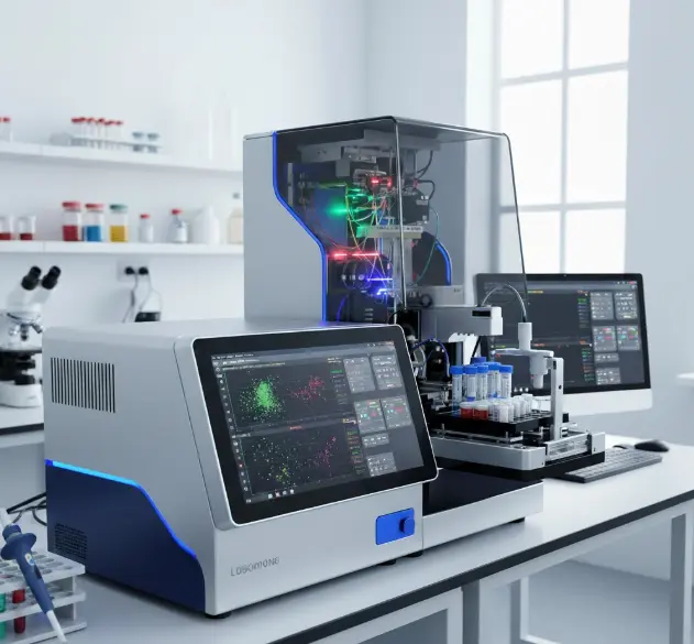
Multicolor Flow Cytometry
Our platform employs advanced multicolor panels capable of simultaneously assessing surface lineage markers, extracellular and intracellular activation markers, and cytokine production profiles at the single-cell level.
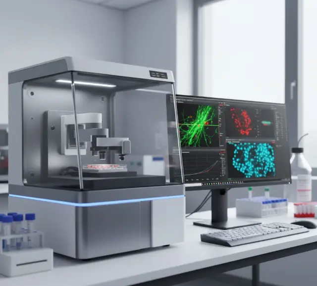
Live-Cell Imaging
We implement sophisticated live-cell imaging techniques to monitor calcium signaling dynamics in real-time, a critical early event in T cell activation that regulates downstream effector functions.
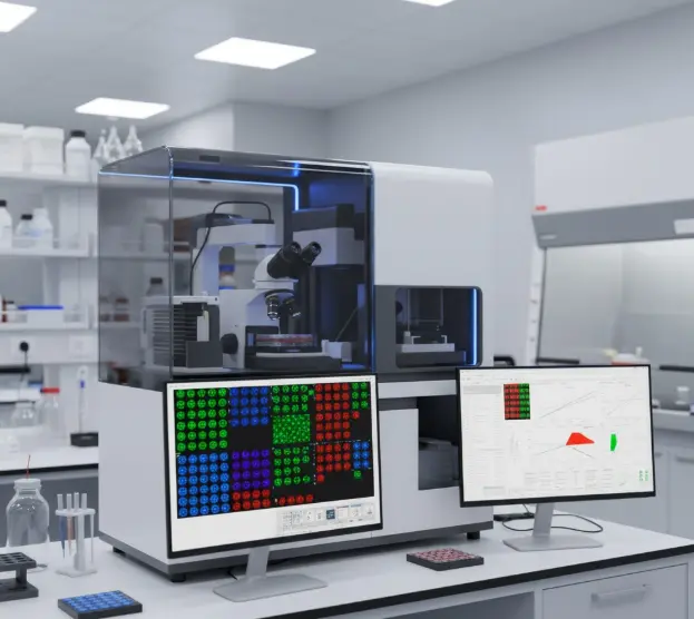
High-Content Screening
The platform incorporates high-throughput capabilities using advanced systems allowing for parallel assessment of multiple activation markers and cytokines in 384-well formats.
T Cell Activation Detection Indicators
Accurate assessment of T cell activation requires monitoring multiple parameters across temporal and functional domains. Our assays capture this complexity through a stratified approach that examines early, intermediate, and late activation events, providing a comprehensive view of the T cell response continuum.
| Marker Category | Specific Markers | Biological Significance | Detection Method |
|---|---|---|---|
| Early Activation | CD69 | Immediate early activation antigen, appears within hours | Flow Cytometry |
| Intermediate Activation | CD25 (IL-2Rα) | Component of high-affinity IL-2 receptor, promotes clonal expansion | Flow Cytometry |
| Late Activation | HLA-DR | MHC Class II molecule indicating prolonged activation | Flow Cytometry |
| Costimulatory/Regulatory | CD279 (PD-1) | Immune checkpoint regulator, exhaustion marker | Flow Cytometry |
| Effector Molecules | Granzyme B | Cytotoxic serine protease | Intracellular Flow Cytometry |
| Metabolic Activation | CD38 | Ectoenzyme with signaling functions, upregulated in activation | Flow Cytometry |
| Cytokine Production | IFN-γ, TNF-α | Key effector cytokines |
Our Services
T cell activation is initiated by the engagement of TCR and CD3 complex, and the subsequent engagement of co-stimulatory molecules, such as CD28 receptor. After co-stimulation of these two signals, a series of intracellular activations happen, including the cytokine release and T cell proliferation in order to fight infection or disease. Scientists at GTOnco™ have developed a systematic approach to T cell activation and proliferation assays for I-O products discovery. The nuclear factor of activated T cells (NFAT), IL-2 or other kinds of factors will be detected by bioluminescent methods or ELISA kits. Meanwhile, the T cell activation and proliferation process can also be evaluated via cell counting kit-8 (CCK-8), MTT, CFSE as well as flow cytometric analysis, etc. We guarantee to provide the best-class service for our clients during the gene therapy-based I-O drugs development.
Why Choose Our Services?
Choosing Creative Biolabs' GTOnco™ platform represents an investment in scientific excellence, data integrity, and regulatory confidence.
Expertise in Gene Therapy I-O
We are not general immunologists and we specialize in gene therapy-based I-O drug development. Our assays are specifically optimized for assessing complex modalities like CAR-T, TCR-T, and oncolytic viruses.
Unrivaled Data Quality
We enforce stringent Quality Control (QC) measures, including post-thaw recovery and viability assessment, and functional validation using standard stimulation protocols, ensuring the functional competence of your cells and the robustness of your data.
Kinetic and Real-Time Analysis
Our integration of live cell imaging and time-course flow cytometry with CFSE allows for the visualization and quantification of real-time phenotype changes and kinetic monitoring of T cell responses, a significant advantage over single endpoint measurements.
Customization Support
From minimum cell requirements to the selection of optional inhibitory and co-stimulatory markers, we offer a fully customizable service backed by in-depth scientific consultation to design the optimal protocol for your specific therapeutic candidate.
Applications
The T Cell Activation & Proliferation Assay is not merely a research tool; it is a gateway to therapeutic validation and development. The data generated by our GTOnco™ platform is directly applicable to major areas of biomedical discovery.
Immune Checkpoint Inhibitor (ICI) Efficacy Evaluation
Determining a compound's ability to reverse T cell anergy/exhaustion by monitoring the upregulation of activation markers (CD25, CD69) and the downregulation of inhibitory receptors (PD-1, CTLA-4) in T cells co-cultured with target cells.
CAR-T and TCR-T Cell Therapy Monitoring
Essential for demonstrating T cell function and persistence in vitro and in vivo . We assess the capacity of genetically engineered T cells to rapidly proliferate and acquire effector function (Granzyme B/Perforin expression) upon contact with their specific tumor antigen.
Cancer Vaccine Response Assessment
Quantifying the magnitude and quality (e.g., Th1 vs. Th2 cytokine profile) of the antigen-specific T cell response induced by a vaccine candidate. This is often done using antigen recall assays in human PBMCs.
Mechanism of Action (MoA) Elucidation
Characterizing the precise immunological pathways affected by novel small molecules or biologics, such as determining if a compound induces T cell activation via a co-stimulatory pathway (CD28, CD137) or modulates intracellular signaling cascades.
Customer Review

"We engaged Creative Biolabs to characterize the activation profile of our novel bispecific T cell engager platform. Their comprehensive analysis included calcium flux measurements alongside traditional activation markers, providing unexpected insights into signaling kinetics that fundamentally improved our candidate selection process. The depth of analysis exceeded our capabilities and accelerated our lead optimization timeline by several months."
Michael Reinhardt, PhD

"For our research on T cell responses in autoimmune inflammation, we needed to track antigen-specific populations across multiple activation parameters. Creative Biolabs' implementation of MHC pentamer technology combined with their extensive polychromatic flow cytometry panels enabled us to identify a rare HLA-DR+CD38hi population that correlated strongly with disease activity. Their technical expertise was instrumental in validating this biomarker cohort."
Sarah Jenkins, PhD

"The GTOnco™ in vivo CFSE proliferation tracking was an absolute game-changer for our studies. We were able to quantitatively demonstrate the superior CD8+ T cell persistence of our novel CAR construct in a humanized mouse model compared to the clinical benchmark. The high-resolution proliferative index provided the critical evidence needed to justify our lead candidate selection and accelerated our project timeline by three months."
Dr. Eleanor Khan
Chief Scientific Officer
Frequently Asked Questions
Our minimum and optimal cell requirements vary based on the assay complexity. For a basic activation analysis, we require a minimum of viable cells per condition. For proliferation assays with CFSE labeling or comprehensive multi-timepoint analyses, we recommend an optimal range of viable cells per condition to ensure robust data and technical replicates.
Absolutely. We offer services to assess T cell specificity via Antigen-specific T cell activation assays using peptide epitope mapping, whole protein antigens, or specific tumor cell lines as targets. This is crucial for evaluating cancer vaccines or TCR-T/CAR-T products.
Yes. Our multiparameter flow cytometry panels always include CD4 and CD8 lineage markers, allowing us to simultaneously analyze and quantify the activation and proliferation of Helper T cells and Cytotoxic T Lymphocytes (CTLs) in the same sample well. This subset-specific analysis is crucial for understanding the therapeutic mechanism.
Yes. Our multiparameter flow cytometry panels always include CD4 and CD8 lineage markers, allowing us to simultaneously analyze and quantify the activation (e.g., CD25+ or CD69+) and proliferation of CD4+ Helper T cells and CD8+ Cytotoxic T Lymphocytes (CTLs) in the same sample well. This subset-specific analysis is crucial for understanding the therapeutic mechanism.
Connect with Us Anytime!
Creative Biolabs is a leader in the field of I-O products discovery with great reputation and experience. We are dedicated to providing professional and advanced in vitro assay platform for our customers around the world. For more detail information, please feel free to contact us.
Start Your Project Today
Tell us about your project, and our experts will get back to you with a customized quote and proposal.