LentiviraL Vector Design for Mitochondria Localization
Creative Biolabs' expertise in vector design and mitochondrial biology, combined with our comprehensive service system, lays a solid foundation for project success. Whether in basic research or drug development, our team is ready to meet your mitochondrial targeting needs with outstanding scientific expertise and technological innovation.
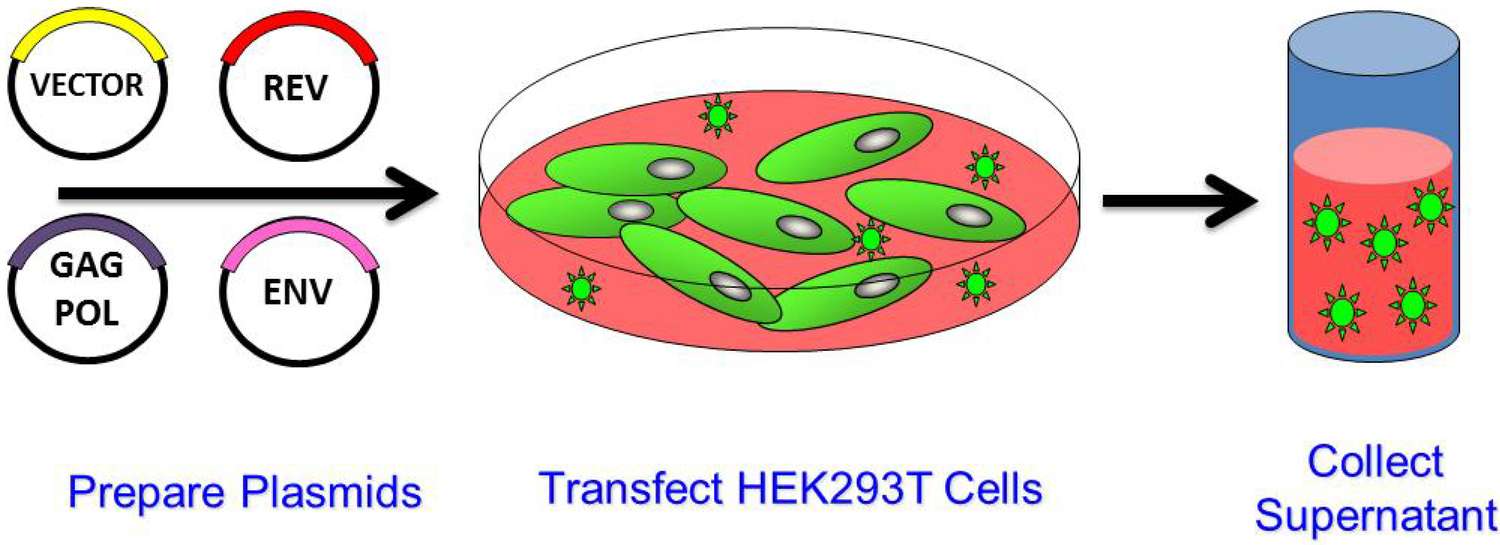 Figure 1. Generation of Lentiviral Vector by Transient Transfection. The four packaging plasmids are transfected into cells that have a high capacity for vector production.1
Overview of Mitochondria Localization
Mitochondrial localization is a technique for studying mitochondrial functioning by targeting specific proteins or compounds in different subregions of the mitochondria. This method involves the fusing of mitochondrial localization signal (MLS) or mitochondrial targeting sequence (MTS) with the target protein or molecule, which allows for precise localization to various compartments inside the mitochondria, such as the matrix, inner membrane, and outer membrane. MTS is a short peptide, 15-70 amino acids long, with positively charged basic residues that drive protein transport to the mitochondria.
The Rationale for Mitochondria Localization in Therapeutics
Mitochondria are far more than just the cell's "powerhouse"; they are a vital signal transduction platform, and their dysfunction is the root cause of a wide range of diseases known as mitochondrial diseases. These diseases range from common conditions like type 2 diabetes and heart failure to rare genetic disorders affecting the central nervous system. Correcting genetic defects or regulating metabolic pathways within this organelle offers unprecedented opportunities for therapeutic intervention.
Principles of lentiviral Vector Design
Basic Structural Components
- Regulatory Elements: Modified long terminal repeats (LTRs) to improve safety and performance
- Packaging Signals: Psi (ψ) sequences crucial for RNA packaging
- Rev Response Elements: Facilitate nuclear export of unspliced RNA
- Marmot Hepatitis Virus Posttranscriptional Regulatory Elements: Enhance transgene expression
- Central Polypurine Sequence: Facilitate nuclear import into non-dividing cells
Safety Engineering Considerations
- Self-Inactivating Vectors: Deletion in the 3' LTR U3 region to inhibit replication
- Isolation Packaging System: Isolates viral genes onto multiple plasmids to prevent recombination
- Tissue-Specific Promoters: Restrict transgene expression to target cell types
- Insulating Elements: Prevent position effects and enhance transgene expression stability
Performance Optimization
- Codon Optimization: Enhance transgene expression through strategic codon usage
- Transcription Optimization: Balance promoter strength and specificity
- Vector Titer Enhancement: Improve viral particle yield and transduction efficiency through modification
- Stability Improvement: Achieve long-term stable expression through engineering modifications
Design of Non-integrating Lentiviral Vectors
Non-integrating lentiviral vectors represent a significant advancement in safety for gene delivery applications. These systems are constructed by specifically mutating integrase, typically involving amino acid substitutions that disrupt catalytic activity while preserving other essential functions. The most common approach utilizes the D64V mutation in the catalytic core domain, which effectively eliminates integration capability while retaining reverse transcription and nuclear importation functions.
Applications and Advantages
Non-integrating lentiviral vectors offer significant advantages in specific therapeutic scenarios:
01.Transient expression requirements: Suitable for applications requiring transient transgenic expression
02.Reduced genotoxicity risk: Eliminates the risk of insertional mutations
03.Stem cell applications: Suitable for the generation and differentiation of induced pluripotent stem cells
04.Gene editing platform: Compatible with nuclease systems
05.Regulated expression systems: Suitable for inducible and tissue-specific expression modalities
Our Services
MTS-based mitochondrial targeting techniques were applied to fusion into viral vectors for targeted delivery. Creative Biolabs created an MTS-conjugated lentiviral vector (LV) that inserts the MTS signal into the transfer plasmid and transfects it into 293T cells using the calcium phosphate technique for lentiviral packaging. We offer several common LVs for mitochondria localization.
- LV-mito-GFP uses green fluorescent protein (GFP) to visualize mitochondria, facilitating dynamic studies of shape and motility.
- LV-mito-mCherry uses the fluorescent protein for multi-channel imaging and colocalization research.
How to Design Lentiviral Vectors for Mitochondria Localization?
Target Signal Selection and Optimization
The design of mitochondrial-targeting lentiviral vectors begins with the strategic selection of suitable target signals. We employ a systematic approach to identify and validate mitochondrial localization sequences from native proteins with mitochondrial tropism. Our platform incorporates sequences derived from cytochrome c oxidase, manganese superoxide dismutase, and ornithine transcarbamylase, among others.
Vector Structure Engineering
The core vector structure integrates multiple functional elements to ensure efficient gene delivery and mitochondrial localization. Our design includes:
- Optimized promoter system for achieving appropriate expression levels
- Strategic localization of the target signal relative to the transgene
- Cleavage sites for proper protein processing
- Regulatory elements for controlling spatiotemporal expression patterns
Mitochondrial Localization Stably Transduced Cell Lines
LV with mitochondrial colocalization is appropriate for establishing stably transduced cell lines. The primary mitochondrial localization stably transduced cell lines we can offer are:
HeLa Cell Line
Stably transfected with the LV-mtDsRed, the HeLa cell line is a fluorescent tool for mitochondrial localization. This fusion protein, which is intended to be localized in mitochondria, produces red fluorescence when illuminated. HeLa cells are widely available and easy to cultivate, making them popular for research on mitochondrial dynamics and functions.
HEK293 Cell Line
Similar to HeLa cells, the HEK293 cell line allows for easy cultivation and transfection. These cells generate green fluorescence when illuminated by mitochondria thanks to stable transfection LV-mito-GFP. This fluorescent allows for high-precision observations of mitochondrial morphology, mobility, and interactions in HEK293 cells.
Neuro-2a Cell Line
The Neuro-2a cell line, also a neuroblastoma cell line, can emit red fluorescence under mitochondrial illumination after transfection of LV-mito-mCherry. It is to be used to study the relationship between mitochondria and neurodegenerative diseases.
Our Collaboration Process: From Concept to Custom Vector
We follow a rigorous, multi-stage collaboration process to ensure your vectors precisely meet your research objectives.
-
Stage 1
In-depth Consultation and Design Specification
The process begins with detailed technical discussions with our PhD-level project leader to identify the therapeutic target, specific mitochondrial subcompartments, required vector titer, and biosafety (integrative or non-integrative). We will finalize the promoter, MTS sequence, and vector backbone structure.
-
Stage 2
Vector Construction and Cloning
We will synthesize a custom expression cassette (including an optimized MTS-transgenic fusion) and carefully clone it into the final lentiviral transfer plasmid backbone. All clones undergo sequence validation to ensure error-free construction.
-
Stage 3
Pilot Production and Validation
We will conduct small-scale pilot production to evaluate vector production efficiency and provide an initial research-grade batch. Crucially, we will perform functional validation assays:
- Transduction efficiency: Measured in the client's target cell lines.
- Location efficiency: Quantitatively confirming the localization of the expressed protein in the target mitochondrial subcompartment using direct immunofluorescence or biochemical separation assays.
-
Phase 4
Mass Production and Quality Assurance
Following the successful completion of pilot validation, we will commence mass production at a certified facility. Rigorous quality control (QC) testing will be conducted at each phase to ensure the final delivery of qualified products.
Frequently Asked Questions
Q: How do you ensure that the expressed protein is localized exclusively to mitochondria?
A: Our assurance is based on a multi-step design and validation process. First, we use proprietary MTS sequences, screened for high affinity interactions with the mitochondrial delivery mechanism (TOM/TIM complex). Second, we conduct rigorous in vitro localization experiments using confocal microscopy and Western blotting of cell lysates. These experiments confirm that the expressed protein is primarily located in the mitochondrial fraction, rather than in the cytoplasm or nucleus, prior to final vector release.
Q: Will your non-integrating lentiviral vector (NILV) undergo integration?
A: While our integrase-deficient mutants, such as the D64V variant, exhibit significantly reduced integration activity (typically over 99%) compared to the wild type, no integrase mutant can achieve 100% non-integration. We validate the effectiveness of NILV using quantitative ALU-PCR to precisely measure residual integration frequency, providing you with clear, quantified safety data that meets the stringent regulatory requirements for transient applications.
Q: What is the optimal promoter choice for mitochondrial-targeted gene delivery?
A: The optimal promoter selection depends entirely on the application. For cell line-based research, constitutive cytomegalovirus (CMV) promoters are typically used to achieve high expression. For in vivo applications, we prioritize tissue-specific promoters (e.g., neuronal, cardiac promoters) or ubiquitous promoters, such as elongation factor-1α (EF1α), to ensure sustained and widespread expression and minimize the risk of adverse immune responses compared to stronger viral promoters. Our consultation phase will tailor a promoter selection strategy to your specific experimental needs.
Q: How do you ensure the stability of the final MTS fusion protein?
A: We employ advanced bioinformatics tools to construct a secondary structure model of the fusion protein. By analyzing the connection region between the MTS and the therapeutic protein, we designed a sequence that minimizes steric hindrance, maximizes the accessibility of the MTS to the delivery mechanism, and ensures that the tertiary structure of the therapeutic domain remains stable and functional after cleavage by mitochondrial processing peptidase (MPP).
Connect with Us Anytime!
Targeted delivery of therapeutic drugs to mitochondria represents a paradigm shift in the treatment of complex diseases. Success in this field requires exceptional expertise in molecular virology, organelle biology, and rigorous quality control. Creative Biolabs is committed to providing high-quality LVs for mitochondrial targeting to customers worldwide. For more information and a quote, please contact us. We will get back to you within 24 hours.
Reference
- Shaw A, Cornetta K. Design and potential of non-integrating lentiviral vectors. Biomedicines, 2014, 2(1): 14-35. https://doi.org/10.3390/biomedicines2010014 (Distributed under Open Access license CC BY 4.0, without modification.)