Jurkat Cell based Cytotoxicity Safety Screening Service
Jurkat Cell Line for Blood Toxicology Studies
Jurkat cells, a human T lymphocyte cell line derived from acute T cell leukemia, are an essential tool for cytotoxicity testing due to their unique properties and versatility in research. Originating from the peripheral blood of a patient with T-cell leukemia, Jurkat cells are characterized by their rapid proliferation and their ability to exhibit many features typical of activated T-cells. These features include the production of interleukin-2 (IL-2) and the expression of key surface antigens relevant to the immune response. Jurkat cells are particularly valuable for blood toxicology studies because they effectively model the human immune response. This makes them exceptionally useful for investigating how various substances impact T-cell function and viability. Their rapid proliferation and sensitivity to toxic agents make them ideal for high-throughput screening, providing crucial insights into the mechanisms of immune response and cytotoxicity.
Jurkat Cell-based Cytotoxicity Screening Service
Understanding the critical need for precise and reliable cytotoxicity assessments, Creative Biolabs proudly offers unmatched Jurkat cell-based cytotoxicity screening services. Our state-of-the-art platform leverages a cell viability assay, a powerful and sensitive method for detecting cytotoxicity, to evaluate the impact of various compounds on Jurkat cells. This assay quantifies ATP levels in Jurkat cells through a luminescent readout, providing a direct and quantitative measure of cell viability. ATP serves as a vital energy carrier in all living cells, with its concentration directly correlating to cell health and viability. Exposure to cytotoxic compounds results in decreased ATP levels, leading to diminished luminescence. By utilizing luminescent reagents that emit a measurable light signal in the presence of ATP, we accurately assess the cytotoxic effects of test compounds on Jurkat cells.
Workflow of Our Service
Creative Biolabs offers a streamlined service workflow for Jurkat cell-based cytotoxicity screening:
- Consultation and Customization: Understanding client-specific needs and customizing the assay setup accordingly.
- Preparation and Assay Setup: Culturing Jurkat cells, preparing test compounds, and setting up the cell viability assay.
- Incubation and Treatment: Exposing Jurkat cells to the test compounds under controlled conditions.
- Luminescence Measurement: Using luminescent readout to assess ATP levels post-treatment.
- Data Analysis: Compiling and analyzing the luminescence data to determine cytotoxic effects.
- Report Generation: Providing clients with detailed reports including raw data, analyzed results, and expert interpretation.
Why Use Luminescent Readout System?
The luminescent readout system offers several advantages over traditional colorimetric methods. It boasts higher sensitivity, enabling the detection of even minimal changes in ATP levels. Additionally, the linearity of the luminescent signal across a broad range of ATP concentrations facilitates accurate quantification, making the detection of subtle cytotoxic effects possible. This methodology thus ensures precision and reliability in our cytotoxicity data.
Published Data
The study examined the cytotoxicity of various nanoparticle formulations, including free DOX, MSNs and RMSNs, to determine cell viability in Jurkat cells. It was observed that free DOX exhibited significantly stronger cytotoxicity toward Jurkat cells compared to DMSNs and RDMSNs. These results suggested a potential for these nanoparticles to reduce side effects.
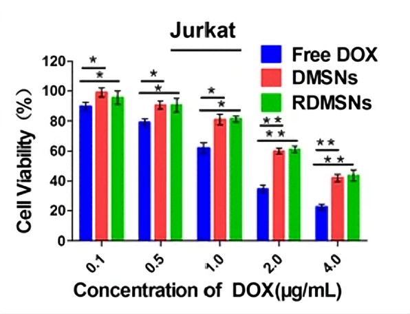 Fig.1 Cell viability study of drug-loaded nanoparticles on Jurkat cells.1,2
Benefits of Our Service
- Suitable for various compound classes
- High-throughput screening
- Sensitivity
- Rapid turnaround time
- Detailed reporting
By choosing Creative Biolabs for your Jurkat cell-based cytotoxicity screening needs, you benefit from our expert team, cutting-edge technology, and commitment to delivering high-quality, actionable data. Contact us today to learn more about how our services can support your research and development efforts.
References
- Zhou, Shoubing, et al. "Intracellular pH-responsive and rituximab-conjugated mesoporous silica nanoparticles for targeted drug delivery to lymphoma B cells." Journal of Experimental & Clinical Cancer Research 36 (2017): 1-14.
- Distributed under Open Access license CC BY 4.0, without modification.
 Download our brochure