

NAA Services for Anti-Insulinoma-Associated-2 (IA-2)
Creative Biolabs is glad to provide natural autoantibodies (NAA) detection and analysis services for our client all over the world. With the consistent growth of various immune diseases (such as type 1 diabetes), it is important for improving the accuracy of diagnosis with suitable methods. We have developed a remarkable and stable platform for NAA services which would be helpful for your diseases diagnosis and treatment projects.
Background of Anti-Insulinoma-Associated-2 (IA-2)
Insulinoma-associated protein-2 (IA-2), also referred to as islet antigen-2 or ICA-512, is a primary autoantigen in type 1 diabetes (T1D). It was found in the nervous tissues and cells of the pancreatic islets and is a member of the protein tyrosine phosphatase family localized to the membrane of dense-core vesicles (DCVs) in neurons and peptide-secreting endocrine cells, including pancreatic β-cells. IA-2 not only contains a transmembrane domain, but also a single catalytic domain of intracellular domain. It is very conservative among species, and its homologues have been identified in various species. Its paralog, a related protein-tyrosine phosphatase (PTP)-like molecule termed IA-2β or phogrin, is also a major islet autoantigen whose location and intracellular domain are 74% identical to IA-2. Among children and adolescents diagnosed with T1D, up to 80% of children and adolescents have anti-IA-2 autoantibody (IA-2 Ab), which commonly develops later in the process causing T1D and is therefore related to more rapid progression.
The Role of Anti-Insulinoma-Associated-2 (IA-2) in Type 1 Diabetes
T1D is a chronic autoimmune disease, which results in severe loss of pancreatic β-cells due to targeting islet cell autoantigens. Studies showed that both IA-2 and IA-2β proteins exhibited calcium-dependent phosphorylation under the action of secretory factors, and IA-2 interacted with cytoskeleton proteins of plasma membrane cells. In IA-2 deficient mice, only 50-60% of the normal insulin secreted from isolated islets was stimulated by glucose in vitro. In addition, blood sugar levels increased significantly in the glucose tolerance test. These results suggest that IA-2 family members play an important role in regulating insulin secretion. Autoantibodies to IA-2 are widely present in T1D patients, providing diagnostic information for diseases, usually before the onset of clinical symptoms. After the cloning of IA-2, the IA-2 serves as an antigen and becomes readily available and methods to detect the autoantibodies and facilitate the routine analysis of large numbers of serum samples.
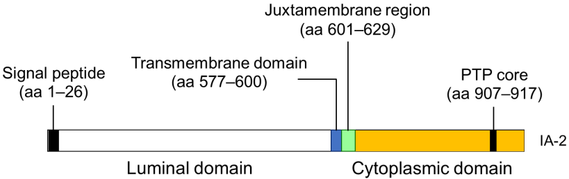 Fig.1 Illustration of antigenic epitopes recognized by T1D sera in IA-2.1
What We Can Do about NAA?
We are able to offer a variety of NAA services with our first-rate technology platforms and prominent scientists, including NAA detection, NAA profiling, NAA affinity measurement, NAA epitope mapping, and paratope mapping. A wide range of NAA products is available for your choice.
Features of Our Services
- First-rate platform
- Experienced scientist
- High efficiency and timely
- Cost-effective service
With our well-established NAA detection and analysis platforms, the professional scientists at Creative Biolabs are dedicated to helping you analyze NAA to IA-2 marker using various customized immunochemical detection methods such as western blotting and ELISA in a timely and cost-effective manner. Our high-quality products and services will greatly contribute to the success of your projects. Please feel free to contact us for more information and a detailed quote.
Reference
- Kawasaki, Eiji. "Anti-Islet Autoantibodies in Type 1 Diabetes." International Journal of Molecular Sciences 24.12 (2023): 10012.
Related Services:
- NAA Services for Anti-GAD65
- NAA Services for Anti-Islet Cell Cytoplasmic
- NAA Services for Anti-Insulin