

Targeted agents can bind to unique tumor biomarkers (e.g., folate receptors, CD105, HER2) or elements of the tumor microenvironment. This application is crucial for the early detection of small lesions, defining tumor margins pre-surgery, monitoring metastatic spread, and accurately staging the disease.
Targeting Delivery for In Vivo Imaging
In the pursuit of precise diagnostics and next-generation theranostics, the ability to visualize molecular events in vivo with high sensitivity is paramount. Our Targeted Delivery Platforms help you accelerate the non-invasive visualization of disease progression and therapeutic response through advanced nanocarrier design, precise surface functionalization, and multimodal probe incorporation. This ensures your imaging agents reach their intended targets efficiently, enhancing diagnostic contrast and enabling real-time monitoring of biological processes.
Click Here to View more about our Service
Targeted Delivery for In vivo Imaging
Targeted Delivery (TD) in the context of in vivo imaging refers to the use of molecular carriers to guide imaging contrast agents or probes to a specific cellular or molecular target within a living organism. This approach moves beyond conventional small-molecule imaging agents, which rely solely on diffusion and natural clearance, often resulting in poor signal-to-noise ratios.
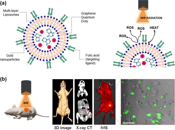 Fig.1 Liposomal nanotheranostics for multimode-targeted bioimaging and phototriggered cancer therapy.1
The core of this technology is the utilization of nanoscale carriers—typically ranging from 10 nm to 200 nm—including liposomes, micelles, dendrimers, and various inorganic nanoparticles (e.g., iron oxide, gold). These nanocarriers serve as highly versatile platforms:
- Structure and Function: They possess a high surface-area-to-volume ratio, allowing for the incorporation of a significant payload of imaging agents, effectively amplifying the signal. Their inner core can encapsulate hydrophobic agents, while their surface can be modified for stability and active targeting.
- Credibility & Mechanism: Cited literature consistently demonstrates that nanocarrier systems address the physicochemical limitations of free probes, improving solubility and protecting them from enzymatic degradation. The ability to modify the surface with ligands such as antibodies (e.g., targeting uPAR on breast cancer cells) drastically improves target-to-background signal ratios, a critical factor for early disease detection and monitoring. Furthermore, the integration of both diagnostic and therapeutic agents within a single nanoparticle defines the emerging and highly promising field of theranostics.
By achieving selective accumulation through either passive (EPR effect) or active targeting, TD nanoplatforms dramatically increase the image contrast, offering unprecedented molecular detail for disease investigation.
Targeted delivery systems primarily focus on two crucial areas of imaging agent development:
-
Fluorescent Imaging Probes
These systems involve synthesizing highly stable, bright fluorophores (often Near-Infrared or NIR dyes for better tissue penetration) and coupling them to high-affinity targeting modules. This approach is highly valued for its non-invasiveness and real-time visualization capabilities, especially in guiding surgical procedures or deep-tissue microscopic analysis. -
Contrast agent delivery
This encompasses the encapsulation or conjugation of conventional agents (such as Gd-chelates for MRI or iodine-based agents for CT) within or onto nanocarriers. The resulting vehicle often exhibits superior T1 or T2 relaxivity compared to the free agent, improving contrast and enabling longer circulation times for enhanced targeting.
Key Applications of Targeted Imaging Nanoplatforms
The ability to precisely position imaging agents opens up vast opportunities across numerous biomedical disciplines. Our targeted imaging platforms are engineered to support applications in both preclinical development and potential clinical translation:
Oncology (Tumor Visualization and Staging)
Theranostics (Image-Guided Therapy)
This represents a combined approach where the nanocarrier simultaneously delivers a therapeutic payload (drug, gene, or heat agent) and an imaging probe. Real-time imaging tracks the carrier's journey to the tumor and monitors the resulting therapeutic effect, providing instantaneous feedback on treatment efficacy and guiding personalized dosing.
Inflammatory and Cardiovascular Disease Assessment
Targeted delivery systems can home in on specific cellular markers of inflammation, such as adhesion molecules or macrophage receptors, providing high-resolution images of atherosclerotic plaques, arthritic joints, or sites of myocardial infarction. This allows for molecular-level assessment of disease activity long before structural changes occur.
Stem Cell Tracking and Gene Therapy Monitoring
For regenerative medicine, nanoprobes can be internalized by transplanted stem cells ex vivo and then tracked in vivo using MRI or optical imaging to confirm their migration, viability, and ultimate integration into the host tissue. Similarly, targeted delivery vehicles loaded with genetic material can be monitored to confirm successful tissue-specific transfection.
What We Can Offer: Comprehensive Imaging Delivery Solutions
Accurate non-invasive imaging often faces hurdles stemming from rapid probe clearance, low signal-to-noise ratios, and non-specific accumulation in off-target organs (like the liver and spleen). Creative Biolabs specializes in overcoming these pharmacokinetic limitations by engineering sophisticated delivery vehicles that direct imaging agents precisely to the diseased site.
Specific Deliverables and Solutions:
Enhanced Contrast and Specificity
We design actively targeted nanocarriers (e.g., liposomes, polymeric nanoparticles, inorganic NPs) functionalized with ligands (peptides, antibodies, aptamers) that specifically bind to overexpressed receptors or biomarkers on target cells (e.g., tumor vasculature, immune cells). This dramatically increases the local concentration of the imaging probe, yielding superior contrast and diagnostic sensitivity compared to passively-delivered agents.
Prolonged Circulation Time
By utilizing stealth coatings, particularly polyethylene glycol (PEGylation), we extend the systemic circulation half-life of your imaging agents, allowing for maximum passive accumulation via the Enhanced Permeability and Retention (EPR) effect in leaky vasculature (common in tumors and inflammation).
Multimodal Integration
We develop integrated platforms capable of carrying multiple imaging probes (e.g., an MRI contrast agent and a near-infrared fluorophore) within a single construct. This enables simultaneous multi-modal imaging, leveraging the anatomical detail of one modality (like MRI) and the high sensitivity of another (like Fluorescence or PET).
Pharmacokinetic and Biodistribution Profiling
We provide comprehensive in vivo studies to track the real-time fate of your targeted imaging agent, offering crucial data on accumulation kinetics, residence time, and clearance pathways necessary for regulatory submissions.
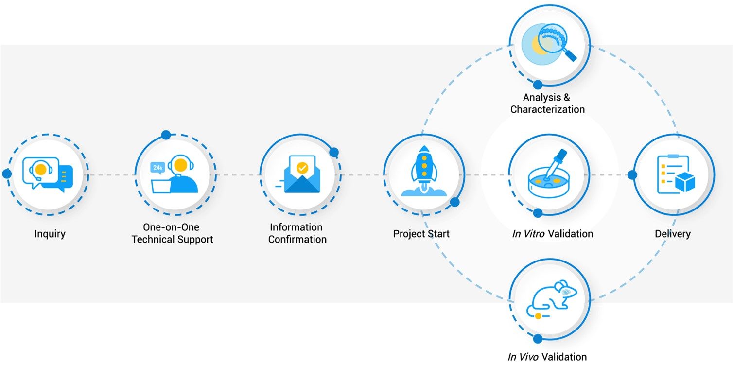
Workflow
FAQs
My current fluorescent probe loses signal rapidly in vivo. How can a delivery system prevent this degradation and background noise?
Nanocarrier encapsulation provides a robust protective shell, shielding the sensitive probe (like a fluorophore or radiotracer) from enzymatic breakdown and rapid clearance by the reticuloendothelial system (RES). Furthermore, by actively targeting the nanocarrier, you concentrate the signal at the site of interest, drastically improving the target-to-background ratio and contrast over time.
We are looking for a system that can both image and deliver a drug. What is the biggest hurdle in combining these functions, and how is it addressed?
The primary challenge is maintaining optimal function for both components simultaneously, as imaging agents and therapeutics often require different environments. The solution involves sophisticated core-shell or multi-layer nanostructures that encapsulate the therapeutic in the core for protection and integrate the imaging probe on the surface or within the shell, ensuring that neither payload interferes with the function or stability of the other.
How do you determine the best targeting ligand for a specific disease, such as a highly heterogeneous tumor type?
Selection is based on expression analysis of surface receptors known to be overexpressed in the target tissue compared to normal tissue. For heterogeneous targets, it is often more effective to target the shared elements of the tumor microenvironment (TME), such as the tumor vasculature or matrix components, using established ligands like RGD peptides or antibodies against CD105, which provides a more consistent target across different cell phenotypes.
What is the optimal particle size range for an actively targeted nanocarrier, considering both tissue penetration and retention?
Generally, particles for solid tumors are often designed to be less than 200 nm to facilitate passive accumulation via the EPR effect, with an ideal size typically falling between 50 nm and 120 nm. Crucially, the size must be large enough to minimize renal filtration (reducing non-specific clearance) but small enough to extravasate through the leaky endothelial barriers present in the target tissue.
Are targeted delivery systems restricted to a single imaging technique, or can the same nanocarrier be adapted for different modalities (like MRI and PET)?
The platform itself is highly versatile. By utilizing a core nanocarrier—such as a mesoporous silica particle or a lipid nanoparticle—you can functionalize it with multiple probes. For example, the same nanoparticle backbone can be conjugated with a chelate for a PET radiotracer and simultaneously loaded with a superparamagnetic iron oxide (SPIO) agent for MRI contrast, creating a powerful, single-injection multimodal imaging tool.
Creative Biolabs provides a crucial partnership for researchers navigating the complexities of in vivo imaging. We specialize in the rational design and synthesis of customized nanoplatforms (liposomes, polymeric NPs, inorganic carriers) that ensure your imaging agents achieve unprecedented target specificity and signal amplification. Our commitment to quality, combined with our expertise in surface chemistry and multimodal integration, transforms challenging diagnostic obstacles into streamlined solutions for disease visualization and theranostic development.
Reference
- Prasad, Rajendra et al. "Liposomal nanotheranostics for multimode targeted in vivo bioimaging and near-infrared light mediated cancer therapy." Communications biology vol. 3,1 284. 5 Jun. 2020, Distributed under Open Access license CC BY 4.0, without modification. https://doi.org/10.1038/s42003-020-1016-z
Our services are For Research Use Only. We do not provide services to individuals.
Online Inquiry