Atomic Force Microscopy (AFM) based Transmembrane Protein Identification Service
Proteins have become an important part of therapeutic drugs or vaccines, and various protein expression systems have thus emerged. In addition to the successful expression of the protein of interest, the identification of protein activity, structure, etc. is also of extreme significance in ensuring the quality of protein. Creative Biolabs has not only established a variety of sophisticated protein expression systems but also developed a downstream protein validation platform.
AFM-based Identification for Transmembrane Protein
AFM uses molecular interactions to identify and measure molecules. Simply put, one molecule attaches to the tip of the AFM and the other molecule attaches to the sample. The specific parameters of the interaction between the molecules on the AFM tip and the molecules in the sample are monitored and recorded, and the relevant information can be inferred by analyzing the final data. An important part of the validation of membrane proteins using AFM is the functionalization of the tip, which involves a series of chemical steps to attach molecules to the tip of the AFM. Membrane proteins surrounding the liposomes can be identified through the recognition of the his-tag by modified tris-Ni+-NTA tip. When performing AFM topographical recording, the AFM tip will circularly approach to the sample and retract from the sample. At each cycle, the cantilever deflection and the distance are monitored and translated into approach and retraction FD curves. Analysis of the results yields the extension of the macromolecule and
Proteins have become an important part of therapeutic drugs or vaccines, and various protein expression systems have thus emerged. In addition to the successful expression of the protein of interest, the identification of protein activity, structure, etc. is also of extreme significance in ensuring the quality of protein. Creative Biolabs has not only established a variety of sophisticated protein expression systems but also developed a downstream protein validation platform.
Background
More than half of the therapeutic targets are membrane proteins, however, producing functional membrane proteins has always been a challenging issue in protein manufacturing. Creative Biolabs' cell-free expression system makes customized protein expression simple and efficient. In order to properly fold the protein, membrane proteins are embedded in the liposome during cell-free protein synthesis. After protein production is completed, validation of the activity and structure of the target protein to ensure protein quality is also an important part of protein production. Atomic force microscopy (AFM) is a nano-scale resolution scanning probe microscope that is 1000 times more powerful than the limits of optical diffraction. It detects the contact between atoms, atomic bonding and so on to present the surface properties of the sample. Unlike scanning electron microscopy, AFM provides a three-dimensional surface map and does not require any special treatment of the sample to avoid irreversible damage to the sample.
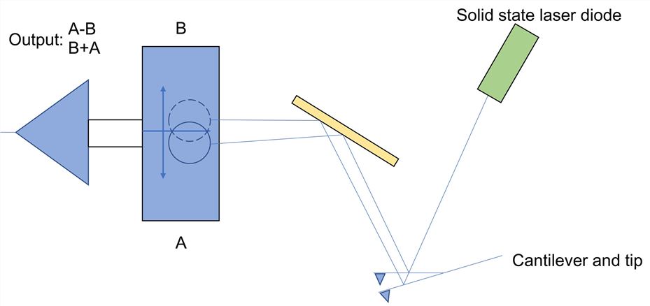 Fig.1 The principle of laser beam reflection detection by atomic force microscope. Distributed under CC BY 3.0, adapted from Wikimedia, without modification.
AFM-based Identification for Transmembrane Protein
AFM uses molecular interactions to identify and measure molecules. Simply put, one molecule attaches to the tip of the AFM and the other molecule attaches to the sample. The specific parameters of the interaction between the molecules on the AFM tip and the molecules in the sample are monitored and recorded, and the relevant information can be inferred by analyzing the final data. An important part of the validation of membrane proteins using AFM is the functionalization of the tip, which involves a series of chemical steps to attach molecules to the tip of the AFM. Membrane proteins surrounding the liposomes can be identified through the recognition of the his-tag by modified tris-Ni+-NTA tip. When performing AFM topographical recording, the AFM tip will circularly approach to the sample and retract from the sample. At each cycle, the cantilever deflection and the distance are monitored and translated into approach and retraction FD curves. Analysis of the results yields the extension of the macromolecule and the number of monomers in the polypeptide.
Advantages of AFM-based Identification for Transmembrane Protein
- High resolution
- Ability to display 3D surface maps
- No special treatment of the sample is required
- Working conditions are simple and easy to implement
The ability of AFM to detect intermolecular interactions by a single molecule is its potential to study the distribution of transmembrane proteins in liposomes. This approach makes validation and quality control of proteins simple. Creative Biolabs not only has a variety of high-quality protein expression systems, but we also carry out rigorous validation and quality control of the proteins we produce, which stems from our establishment of a series of scientific and effective validation methods.
the number of monomers in the polypeptide.
Advantages of AFM-based Identification for Transmembrane Protein
- High resolution
- Ability to display 3D surface maps
- No special treatment of the sample is required
- Working conditions are simple and easy to implement
The ability of AFM to detect intermolecular interactions by a single molecule is its potential to study the distribution of transmembrane proteins in liposomes. This approach makes validation and quality control of proteins simple. Creative Biolabs not only has a variety of high-quality protein expression systems, but we also carry out rigorous validation and quality control of the proteins we produce, which stems from our establishment of a series of scientific and effective validation methods.
All of our products can only be used for research purposes. These vaccine ingredients CANNOT be used directly on humans or animals.
