RSV Infection Life Cycle and Vaccine Target Discovery
Respiratory Syncytial Virus (RSV) is a global health threat, with high risks for infants under six months and adults over 65. It causes millions of lower respiratory tract infections yearly, including bronchiolitis and pneumonia, leading to hospitalizations and deaths. Understanding RSV's infection life cycle is key to finding effective vaccine targets, as each stage offers intervention opportunities. This review breaks down the virus's life cycle, key proteins, host interactions, and vaccine progress for biomedical researchers.
RSV Fundamentals: Classification, Genome, and Structure
Viral Classification and Genomic Organization
RSV belongs to the Pneumoviridae family, Mononegavirales order, and Orthopneumovirus genus. It has two main subtypes (A and B) that differ in the attachment (G) protein, causing antigenic variation and complicating vaccine development.
The RSV genome is ~15 kb, single-stranded negative-sense RNA, encoding 10 major genes. Six are structural proteins (nucleocapsid N, phosphoprotein P, matrix M, fusion F, attachment G, small hydrophobic SH) critical for assembly, entry, and immune recognition. Four are non-structural proteins (NS1, NS2, M2-1, M2-2) that suppress host immunity and regulate replication. The genome is wrapped in N protein to form a ribonucleoprotein (RNP) complex, which, with P (polymerase cofactor) and L (RNA polymerase) proteins, enables transcription and replication.
The RSV Infection Life Cycle: A Step-by-Step Breakdown
Attachment
The G protein mediates viral attachment to host cells via two receptors:
- CX3CR1: G protein's CX3C motif mimics human chemokine CX3CL1, binding to CX3CR1 on respiratory epithelial and immune cells. This aids attachment and disrupts immune signaling.
- GAGs: G protein's heparin-binding domain (HBD) binds negatively charged glycosaminoglycans (e.g., heparan sulfate) on cells, concentrating viruses for better CX3CR1 interaction.
The F protein may also bind secondary receptor nucleolin to assist attachment.
Fusion
F protein drives membrane fusion, switching from prefusion (preF) to postfusion (postF) conformations. PreF is antigenically critical, exposing neutralizing epitopes. Fusion steps:
- PreF binds receptors (e.g., nucleolin).
- Conformational shift reveals a hydrophobic fusion peptide.
- Peptide inserts into host membrane, bringing viral and host membranes close.
- Further shifts fuse membranes, releasing the nucleocapsid into the cytoplasm.
Transcription/Replication
Viral inclusion bodies (IBs) in the cytoplasm—formed by N, P, M2-1 proteins—protect RNA synthesis:
- Transcription: L protein makes positive-sense mRNAs (3′ to 5′ order), capped and polyadenylated for translation. M2-1 ensures complete gene transcription.
- Replication: Once enough N/P proteins form, L protein makes full-length positive-sense cRNA, which templates new negative-sense genomic RNA. New genomes are wrapped in N protein for assembly.
Protein Translation
Viral mRNAs are translated in the cytoplasm. F/G proteins are glycosylated in the ER/Golgi for stability. Non-structural proteins (e.g., NS1) suppress host interferon signaling early.
Assembly
At the plasma membrane, M protein links nucleocapsids to F/G/SH proteins (transported from ER/Golgi). New nucleocapsids bind M protein, and glycoproteins cluster at assembly sites.
Budding
M protein forms membrane buds around assembled components. Virions pinch off, releasing infectious RSV. SH protein may aid budding by modifying membrane fluidity.
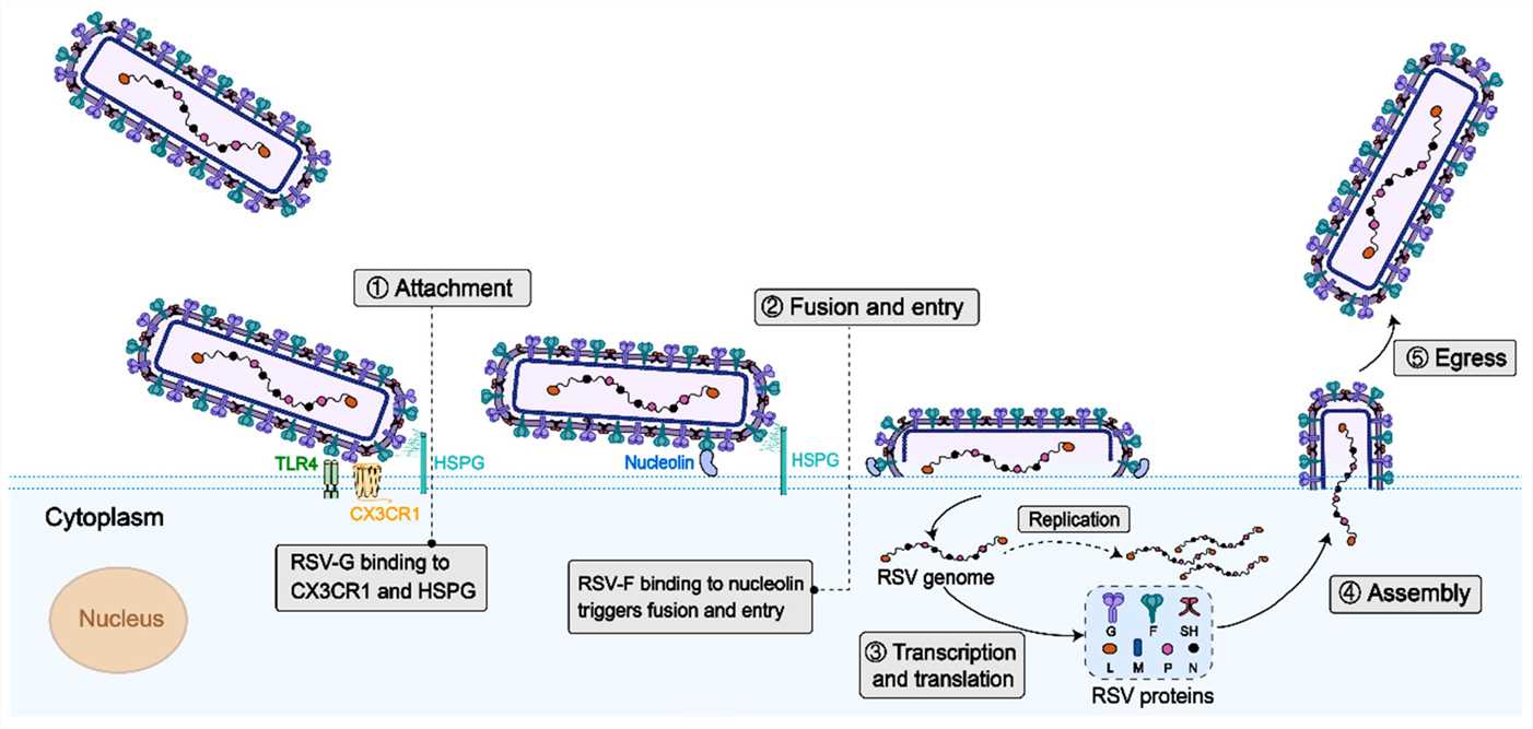 Fig.1 Schematic Illustration of RSV Life Cycle.1,2
Services you may interested in
Critical Surface Glycoproteins and Their Functions
The three surface glycoproteins—F, G, and SH—are central to RSV pathogenesis and are the primary focus for therapeutic development.
F Protein
The F protein is responsible for mediating the critical step of viral-to-cell membrane fusion.
- Key Conformation: The pre-fusion F conformation (Prefusion F) has been identified as the structure possessing the highest activity for inducing neutralizing antibodies.
- Vaccine Significance: The structural resolution of the F protein is a core component of modern vaccine design strategies.
The Attachment (G) Protein
The G protein facilitates the initial attachment of the virus to the host cell.
- Immune Evasion: The G protein contains a central conserved domain (CCD) that includes a CX3C chemokine motif. This motif mimics the host chemokine CX3CL, which is thought to induce immune evasion.
- Cell Binding: The G protein also possesses a Heparin Binding Domain (HBD) which demonstrates high affinity for glycosaminoglycans (GAGs) on the cell surface.
The Small Hydrophobic (SH) Protein
Non-essential in cell culture but modulates host immunity (e.g., inhibiting interferons) in vivo. Including SH in multivalent vaccines (with F/G) induces broader immunity in preclinical tests, though more research is needed.
RSV Vaccine Development Progress
| Platform | Targets | Progress |
|---|---|---|
| Prefusion F Subunit | Stabilized preF | Advanced, safe/efficacious in trials (protects infants/older adults) |
| G Protein Nanoparticles | G's CX3C/CCD | Preclinical success (cross-subtype protection via nanocarriers) |
| mRNA Vaccines | preF or full genome | Ongoing trials (2024–2025); LNPs deliver mRNA, induce strong responses |
| Monoclonal Antibodies | F protein | Prevents high-risk infant infection; long-acting versions in development |
| Multivalent Vaccines | F+G+SH | Early trials (e.g., phase I); broader immunity than single-target |
Challenges and Future Directions
Antibody Escape
F protein mutations can reduce vaccine-induced antibody binding. Solutions: target conserved epitopes (e.g., F's fusion peptide) and monitor global RSV sequences.
Older Adults' Immune Responses
Immunosenescence weakens vaccine responses. Strategies: adjuvants (e.g., TLR agonists), prime-boost regimens, and targeting CD8+ T cells.
Global Accessibility
Cold chain needs and high costs limit access in LMICs. Efforts: thermostable formulations, low-cost manufacturing, and global health partnerships.
Technological Advances
- Cryo-EM reveals new preF epitopes.
- RNA structural biology targets conserved genomic domains for antivirals.
- High-throughput sequencing uncovers host-virus interactions to identify new targets.
Conclusion
The complete life cycle of RSV offers multiple potential intervention points. The comprehensive analysis of the viral structure, particularly the prefusion F protein and the GCX3C motif, provides a strong foundation for vaccine development. By integrating structural biology, studies on RNA secondary structure, and host receptor interactions, the research community can accelerate the development of new, broad-spectrum RSV vaccines and therapeutics. This multi-pronged approach is crucial for addressing the pervasive threat of RSV infection worldwide.
If you want to learn more about the norovirus vaccine, please refer to:
Understanding the Molecular Mechanisms of Three RSV Vaccine Construction Strategies
Respiratory Syncytial Virus F Protein in Next-Generation Vaccine Design
The Translational Value of BRSV Models in Human RSV Vaccine
Deciphering the Molecular Mechanisms of Respiratory Syncytial Virus Infection
Browse our Norovirus Antigen Products
Need a custom solution? If our off-the-shelf products aren't a perfect fit, we can create one for you. Contact us to design a product that precisely matches your experimental demands.
References
- Deng, Lie, et al. "Progress on respiratory syncytial virus vaccine development and evaluation methods." Vaccines 13.3 (2025): 304. https://doi.org/10.3390/vaccines13030304
- Distributed under Open Access license CC BY 4.0, without modification.
Created October 2025
All of our products can only be used for research purposes. These vaccine ingredients CANNOT be used directly on humans or animals.
