Urinary System Disease Model Construction Service for Exosome Functional Research
Overview Disease Model FAQs
Overview
At present, the diagnosis and treatment of most urinary system diseases (USDs) lack specificity and sensitivity, which can easily cause delays in the disease and other unnecessary damage to the body. Therefore, it is important to find better ways to diagnose and treat USDs. In recent years, with the development of targeted therapy technology, exosomes have gradually become an important target for disease diagnosis and treatment due to their good targeting and tolerance. A variety of cells can release exosomes, which are membranous vesicles with the function of carrying substances and transmitting signals, and are the communication media between almost all cells. A large number of studies have shown that natural exosomes or engineered exosomes have a good therapeutic effect on USDs. These findings are inseparable from a suitable USD animal model. Creative Biolabs has been paying attention to the research progress of USD-related exosome therapy and diagnosis and has been updating its technology and knowledge reserves. We can provide customers with the most suitable USD animal model and one-stop services including exosome extraction, exosome identification, exosome engineering, exosome labeling, and in vivo and in vitro verification of exosomes.
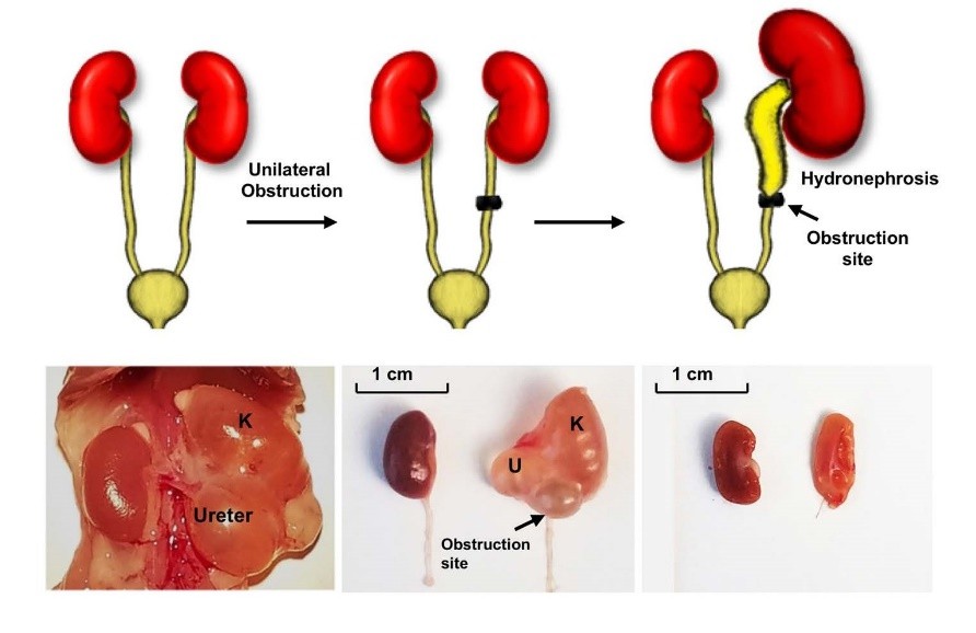 Fig.1 Schema of generation of the Unilateral ureteral obstruction (UUO) model. (Belghasem, 2019)
Creative Biolabs Urinary System Disease Model Library for Exosome Functional Research
We can provide including but not limited to the following USD animal models for exosome functional research.
Creative Biolabs has more efficient experimental facilities and richer project experience and can provide more USD animal models for customers to choose from. Please feel free to contact us with your needs. Our professional team will design a more reasonable experimental plan for you to accelerate the progress of your project.
Reference
-
Belghasem, ME.; A'amar, O.; et al. Towards minimally-invasive, quantitative assessment of chronic kidney disease using optical spectroscopy. Scientific Reports. 2019. 9(1):7168.
FAQs
What is the relevance of exosomes in urinary system diseases, and how can they be utilized for disease modeling?
Exosomes play crucial roles in the pathogenesis of urinary system diseases, including kidney disorders, urinary tract infections, and bladder cancer. Our service leverages exosomes to construct disease models that mimic key pathological features, providing valuable platforms for studying disease mechanisms and evaluating therapeutic strategies.
How are exosomes sourced for urinary system disease model construction, and what advantages do they offer in this context?
Exosomes can be isolated from urine, blood, or kidney tissue, serving as rich sources of disease-specific biomarkers and mediators. Utilizing exosomes for disease modeling offers advantages such as non-invasiveness of sample collection, physiological relevance, and the ability to capture disease-specific molecular signatures, facilitating accurate disease modeling and therapeutic development.
Which urinary system diseases can be modeled using exosomes, and how accurately do these models recapitulate human pathophysiology?
Our service enables the modeling of various urinary system diseases, including chronic kidney disease (CKD), nephrotic syndrome, urinary tract infections (UTIs), and renal cell carcinoma (RCC). These models closely mimic human pathophysiology by incorporating disease-associated exosomes containing biomolecules indicative of disease status, allowing for the study of disease progression and therapeutic responses.
What methodologies are involved in constructing urinary system disease models using exosomes, and what experimental approaches are employed?
Urinary system disease models are constructed by administering disease-associated exosomes to relevant in vitro or in vivo model systems, such as cell cultures or animal models. Experimental approaches include exosome uptake assays, functional assays, histological analysis, and molecular profiling techniques to assess disease-relevant phenotypic changes and therapeutic responses accurately.
Can these exosome-based disease models be tailored to address specific research questions or therapeutic targets?
Yes, our service offers customizable disease models tailored to address specific research questions, therapeutic targets, or experimental requirements. Researchers can choose disease-relevant exosome sources, model systems, disease endpoints, and outcome measures, allowing for flexible study designs and the exploration of diverse research avenues.
For Research Use Only. Cannot be used by patients.
Related Services: