Although Wnt proteins are hydrophobic glycoproteins, they are capable of long-range signaling. Researchers at The Ottawa Hospital Research Institute in Canada discovered that Wnt7a is secreted as extracellular vesicle (EV)-associated proteins after muscle injury. They identified a signal peptide region in Wnts, termed the Exosome Binding Peptide (EBP), which is essential for secretion onto EVs. Adding the EBP to an unrelated protein enabled its secretion via EVs. A di-lysine motif mediates the interaction between the EBP and coat proteins, a function conserved across the Wnt family. This study elucidates the structural basis and uniqueness of Wnt secretion via EVs, distinguishing it from classical secretion pathways. It provides new avenues for innovative therapeutic targeting strategies and systemic protein delivery. The findings were published online on December 13 in the internationally renowned multidisciplinary journal Science Advances, under the title “Identification of the Wnt signal peptide that directs secretion on extracellular vesicles.”
Wnt proteins are a conserved family of secreted glycoproteins responsible for regulating fundamental processes such as development, growth, and regeneration, as well as participating in pathological conditions such as cancer. Wnt signaling plays multiple roles in regulating stem cell functions, including proliferation, cell polarity, symmetric division, motility, and fate determination. Despite their relative hydrophobicity of Wnt proteins due to their palmitoylation, they actively participate in long-range paracrine signaling between Wnt-producing cells and distant receptor cells. Several mechanisms have been proposed to explain long-range Wnt signaling, including transfer via lipoproteins, transport through cellular extensions called cytonemes, binding to soluble Wnt-binding proteins, or packaging into small extracellular vesicles (EVs) known as exosomes.
Exosomes are small (40–150 nm) extracellular vesicles involved in intercellular communication by transferring bioactive cargo, such as lipids, proteins, microRNAs, and mRNAs, to distal cells. Numerous in vitro studies have demonstrated that different Wnt proteins are secreted on the surface of exosomal EVs, and EV-associated Wnt can elicit appropriate signaling in target cells. Substantial in vivo evidence from Caenorhabditis elegans and Drosophila supports the critical role of EVs in long-range Wnt signaling. However, the mechanism by which Wnt associates with these migratory EVs remains unknown.
After acute injury in adult skeletal muscle, Wnt7a is significantly upregulated and actively stimulates regenerative myogenesis at multiple levels. Wnt7a/Fzd7 signaling promotes symmetric muscle stem cell expansion and motility via the planar cell polarity pathway. Additionally, it stimulates anabolic growth and hypertrophy in muscle fibersvia the AKT/mTOR pathway. Intramuscular injection of Wnt7a protein has been shown to significantly improve disease progression in mdx mice, a model for Duchenne muscular dystrophy (DMD). These findings highlight Wnt7a as a promising therapeutic candidate for DMD. However, due to its high hydrophobicity conferred by conserved palmitoylation, delivering Wnt7a through the circulatory system remains a challenge. Therefore, researchers have investigated whether Wnt7a is secreted via EVs and have sought to elucidate the mechanisms responsible for its EV loading, aiming to provide insights into long-range Wnt signaling for treating neuromuscular diseases.
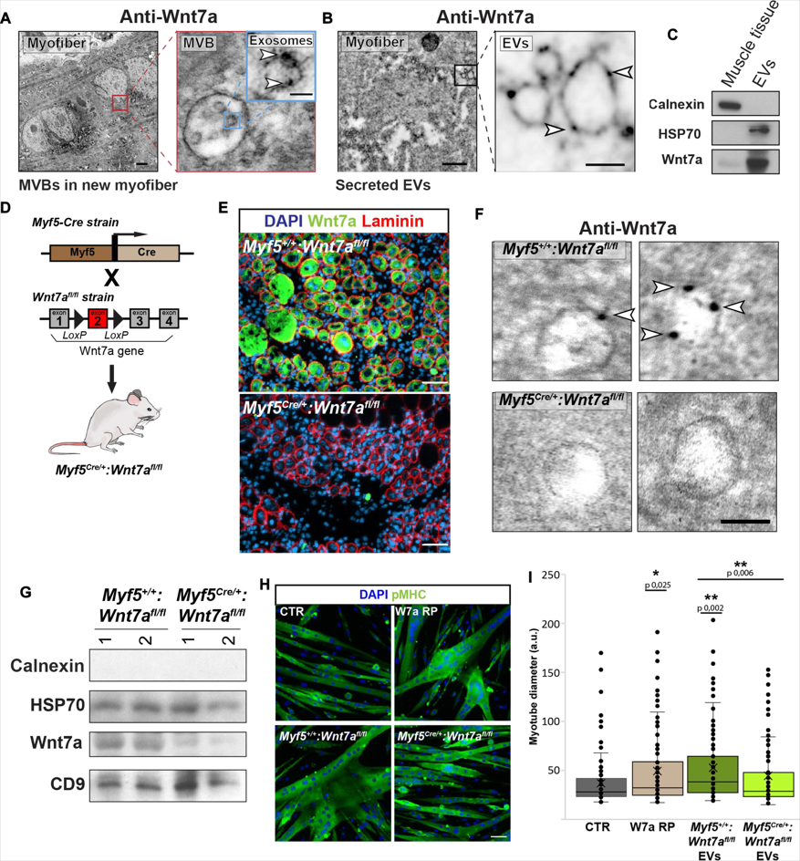
The researchers conducted a structure-function deletion analysis and identified an 18-amino acid signal sequence in Wnt7a, termed the exosome-binding peptide (EBP). Notably, linking the EBP sequence to other proteins resulted in their secretion onto EVs. Surprisingly, neither palmitoylation nor the N-terminal signal peptide (SP) was required for Wnt7a secretion on EVs. Knockdown of WLS abolished the secretion of non-EV-associated Wnt7a but only partially reduced Wnt7a-EV secretion. Through BioID analysis, the researchers identified coat proteins COPA and COPB2 as essential for EBP binding and for localizing Wnt7a to the EV surface.
Finally, co-crystal structural analysis, isothermal titration calorimetry (ITC), and mutational studies revealed a direct interaction between the positively charged di-lysine motif (KIK) in EBP and COPB2. Disrupting the interaction between Wnt7a and the coat proteins impaired the muscle regeneration response in vivo. The researchers provided evidence suggesting that this mechanism is conserved across the Wnt family. This discovery elucidates a non-classical structural mechanism mediating Wnt secretion on EVs and offers insights into the uniqueness of long-range Wnt-EV signaling.
Reference:
Gurriaran-Rodriguez, Uxia et al. “Identification of the Wnt signal peptide that directs secretion on extracellular vesicles.” Science Advances vol. 10,50 (2024): eado5914. doi:10.1126/sciadv.ado5914
Related Services: