There is no doubt that the emergence of immunotherapy has redefined the field of cancer treatment as breakthrough progress. Most immunotherapies now focus on the adaptive immune response, that is, on T cells. Of course, T cells are also very competitive with excellent effect in either CAR-T therapy or immunological checkpoint inhibitors led by PD-1/PD-L1.
Cunning cancer cells are not so easily to be recognized by T cells. However, in our immune system, natural killer (NK) cells also play a major role to kill cancer cells with prior effects than T cells in the innate immune response.
The innate immune response is the first defense line of the immune system, which is not specific without requiring complicated antigen presentation. Therefore, compared with T cells, NK cells possess more effective killing ability with a wider attacking scope.
In the recent Science, researchers at the Dana-Farber Cancer Institute at Harvard Medical School screened a monoclonal antibody that enhanced the ability of NK cells to recognize and kill cancer cells. [1] With the commentary published in the same period, titled Natural killers join the fight against cancer, this study expands the scope of immunotherapy beyond T cells [2].
Let’s briefly look at how the researchers did it. MICA/MICB (MHCI-related protein A/B) are a pair of proteins on the surface of cancer cells, which express the signal and are identified by NKG2D, the NK cell surface receptor. Then NK cells release perforin and granzymes to dissolve cancer cells or induce cancer cell apoptosis, thereby killing cancer cells.
MICA/MICB is expressed under genomic lesions and cellular stress. And it is also expressed on the surface of various types of cancer cells such as melanoma, lung cancer, and leukemia. They are also the first ligands to bind to NKG2D receptors.
Since cancer cells can evade the attack of T cells, naturally, they can also find a way to avoid NK cells. They release various enzymes to hydrolyze these two proteins and let them fall off the cell surface. In this case, without the ligand binding to the NKG2D receptor, NK cells will not kill cancer cells.
So researchers think that MICA/MICB can be firmly attached to the surface of the cancer cells to recruit NK cells to kill tumors. In this way, they screened the monoclonal antibody called 7C6.
According to the previous study of protein structure, the domains of MICA and MICB can be divided into three parts, where α1 and α2 are responsible for binding with NKG2D, while α3 is responsible for adhesion to the surface of cancer cells, so α3 becomes the target where they are hydrolyzed and fall off. Therefore, this mAb specifically binds to the α3 domain without affecting the binding of α1 and α2 to NKG2D.
They designed three monoclonal antibodies and examined them one by one in several cancer cell lines. Then they found that 7C6 not only had the best binding effect but also enhanced the ability of NK cells to kill cancer cells.
In a mouse model of melanoma and colon cancer with lung metastases, 7C6 not only controlled the primary tumor but also visually eliminated the metastases that had metastasized to the lungs. Correspondingly, the researchers barely detected shedding MICA and MICB. In addition, they also transferred human melanoma cells to immunodeficient mice and injected them with human NK cells and cytokines that support the survival of NK cells, interleukin-2.
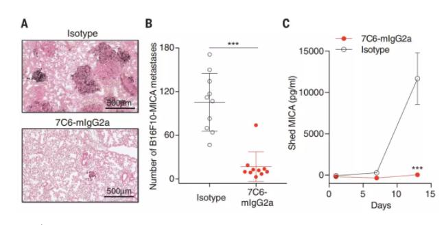
Melanoma tumor lung metastasis model: lung tissue slices (A), number of lung metastases (B), and shedding of MICA (C) after treatment with 7C6 monoclonal antibody (7C6-mlgG2a) and irrelevant isotype in the control group situation
The tumor metastasis of these mice not only remains in lung, but also in liver, pancreas, kidney, adrenal gland and brain, in which liver metastasis is especially obvious, causing severe liver damage. Subsequently, the researchers observed that the injection of 7C6 mAb and NK cells did well in eliminating metastases. But it was a little different in the liver because they found that metastasis could be largely cleared even if they only injected 7C6 mAb. NK cells could make the removal more thorough as icing on the cake.
Through investigation, the researchers found that the injection of 7C6 mAb activated the Kupffer cell. It is a specialized macrophage in the liver equipped with the same phagocytic function as ordinary macrophages and it also has receptors that recognize MICA/MICB to bring a particularly good effect in cleaning metastatic cancer cells. Given that MICA/MICB is present in many solid tumors and hematological cancers, 7C6 mAb therapy will have a wide range of applications.
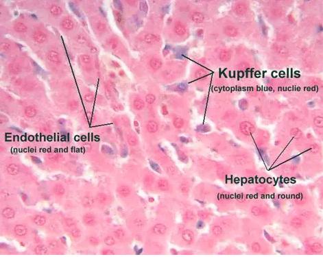
Liver tissue staining sections: endothelial cells (left), Kupffer cells (middle), and hepatocytes (right)
In the review article, two commentators pointed out that the expression of MICA and MICB genes is very complex with many variants, and the binding ability to NKG2D receptors is also not the same. In this new study, 7C6 mAbs are targeting the most common MICA/MICB variants, which are expressed on many tumor cells but are virtually absent on the surface of healthy cells.
In addition to NK cells, the NKG2D receptor is also expressed on the surface of CD8+ T cells. That is, when the NKG2D receptor pathway is activated, CD8+ T cells can also be activated.
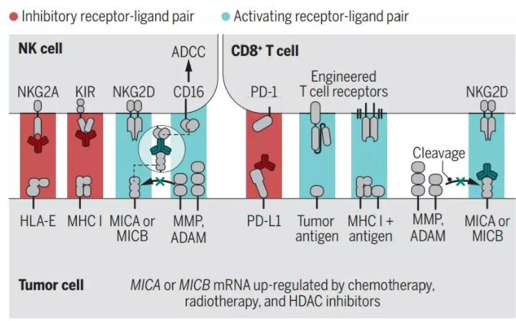
Different pathways associated with NK cells and CD8+ T cells: red is the inhibitory pathway and blue is the activation pathway. NKG2D pathway is active in both cells
Although in this new study, the researchers found that the elimination of tumor cells caused by the use of 7C6 monoclonal antibody is mainly mediated by NK cells, but two commentators believe that more studies are needed to verify whether or not CD8+ T cells also contribute to them, and this also offers the possibility of combination treatment of MICA/MICB monoclonal antibodies and immune checkpoint inhibitors or other T cell immunotherapy.
Commentators at the University of California pointed out that compared with traditional therapies that directly attack tumors, new therapies that target the immune system against tumors are better choices. For patients, there will be fewer side effects and a better prognosis, and that’s why they’re getting more and more attention.
References:
[1] de Andrade L F, Tay R E, Pan D, et al. Antibody-mediated inhibition of MICA and MICB shedding promotes NK cell-driven tumor immunity[J]. Science, 2018, 359(6383): 1537-1542.
[2] Cerwenka A, Lanier L L. Natural killers join the fight against cancer[J]. Science, 2018, 359(6383): 1460-1461.
[3] Wang X, Lundgren AD, Singh P, et al. An six-amino acid motif in the α3 domain of MICA is the cancer therapeutic target to inhibit shedding[J]. Biochemical and biophysical research communications, 2009, 387(3) ): 476-481.
[4] Li P, Morris D L, Willcox B E, et al. Complex structure of the activating immunoreceptor NKG2D and its MHC class I–like ligand MICA[J]. Nature immunology, 2001, 2(5): 443.
[5] Roberts AI, Lee L, Schwarz E, et al. Cutting edge: NKG2D receptors induced by IL-15 costimulate CD28-negative effector CTL in the tissue microenvironment[J]. The Journal of Immunology, 2001, 167(10) : 5527-5530.
[6] https://medicalxpress.com/news/2018-03-tumors-proteins-immune.html