Extracellular vesicles (EVs)are a type of vesicle secreted by cells, characterized by a phospholipid bilayer structure. They carry a wealth of molecular information, including specific proteins, nucleic acids, lipids, and metabolites from the parent cell. Widely present in body fluids, EVs have emerged as crucial biomarkers for disease diagnosis and treatment evaluation in liquid biopsy technology. However, current methods for the rapid purification and detection of extracellular vesicles face challenges such as complex body fluid sample purification, tedious labeling steps, and difficulties in signal readout, indicating the need for further development.
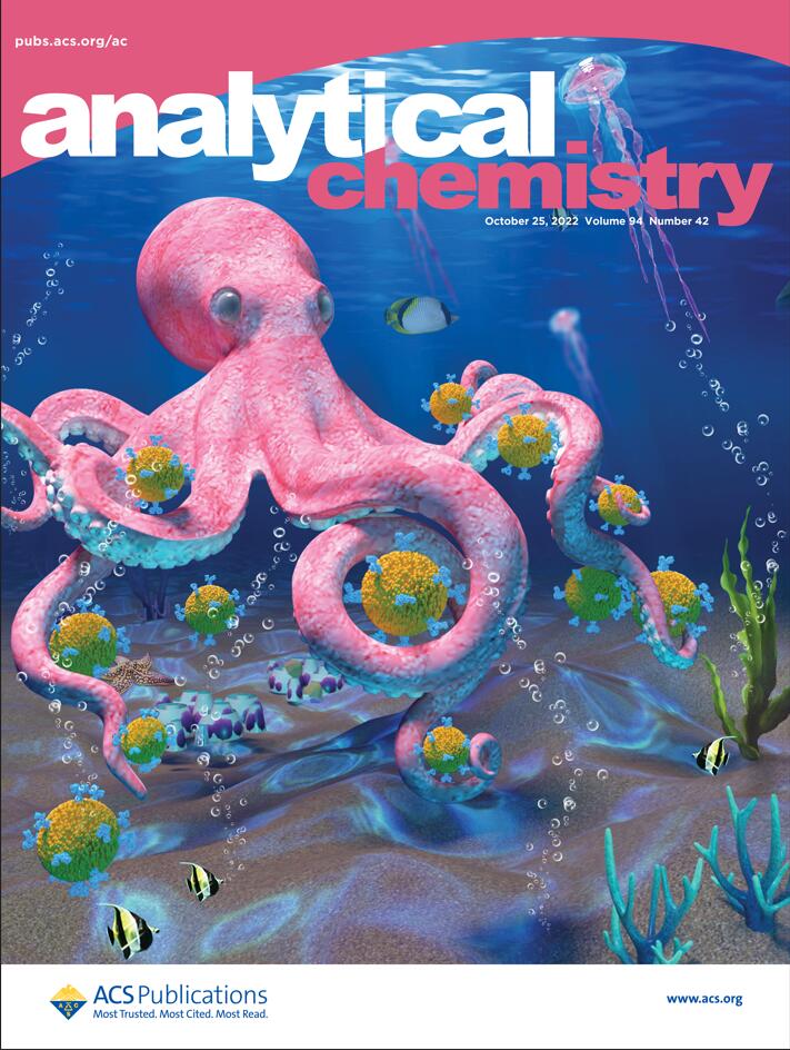
Recently, Analytical Chemistry published a research paper titled “Biomimetic 3D Recognition with 2D Flexible Nanoarchitectures for Ultrasensitive and Visual Extracellular Vesicle Detection,” which was featured as the cover of the current issue.
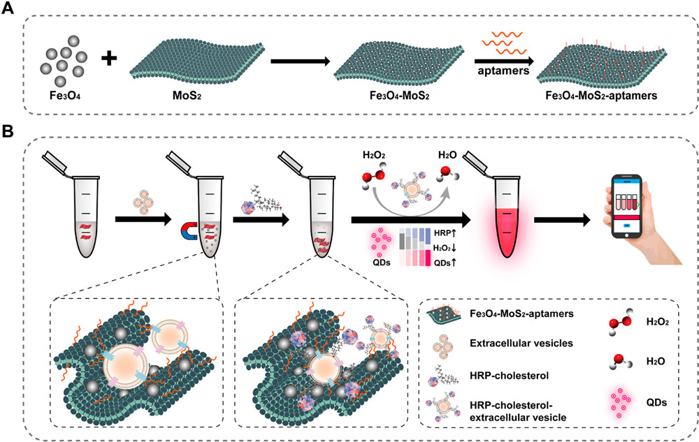
The research team introduced a high-affinity recognition and visual extracellular vesicle detection method named “HARVEST,” utilizing two-dimensional flexible Fe3O4-MoS2 nanostructures. Drawing inspiration from octopus predatory behavior, this method employs spatial recognition and multi-dental combination to biomimetically achieve three-dimensional recognition and capture of extracellular vesicles, significantly enhancing capture efficiency.
This work integrates two-dimensional flexible Fe3O4-MoS2 capture nanostructures with cholesterol lipid labeling chemistry, facilitating extracellular vesicle signal detection through the introduction of a fluorescence visualization system. Specifically, magnetic nanoparticles Fe3O4 and tumor marker aptamers are introduced into two-dimensional MoS2 for magnetic-specific capture of tumor-derived extracellular vesicles. Subsequently, HRP-cholesterol is anchored on the extracellular vesicle surface for lipid labeling, forming an HRP-EV complex. Within this complex, HRP catalyzes the decomposition of H2O2, causing an increase in fluorescence intensity of subsequently added H2O2-sensitive fluorescent quantum dots (QDs) as the concentration of HRP-EV rises. Further quantification of fluorescence values through smartphones enables the analysis of extracellular vesicle marker levels, converting biological signals into optical signals in the “extracellular vesicle-cholesterol-QD” system.
This study establishes an efficient and convenient platform for the rapid enrichment and visual detection of extracellular vesicles. The high specific surface area and mechanical flexibility of aptamer-functionalized two-dimensional platforms enhance aptamer recognition, resulting in a higher capture rate of extracellular vesicles in a shorter time. This underscores its potential for sensitive and accurate detection of circulating biomarkers. In the current study, only the single extracellular vesicle marker CD44 was selected for clinical sample diagnosis. Subsequent detection can be expanded through the combination of multiple markers to further enhance diagnostic accuracy and broaden clinical applications.
Reference:
Li Z, Ma D, Zhang Y, et al. Biomimetic 3D Recognition with 2D Flexible Nanoarchitectures for Ultrasensitive and Visual Extracellular Vesicle Detection. Anal Chem. 2022;94(42):14794-14800. doi:10.1021/acs.analchem.2c03839
Related Services: