Venom Peptide Ion Channel Screening Service
Ion Channels
Ion channels have a very important functional role in immunotherapy. Among them, blocking voltage-gated potassium (Kv) channels have been shown to inhibit immune activation. In addition, ligand-gated ion channels such as the niacin acetylcholine receptor (nAChR) also play a minor role in the immune system. Venom peptides, such as conotoxins, have been shown to exhibit diversity and specificity for membrane protein targets. Pharmacological targets associated with venom peptides include voltage-gated potassium channels, voltage-gated calcium channels (Cav), nicotinic and acetylcholine receptors, among others.
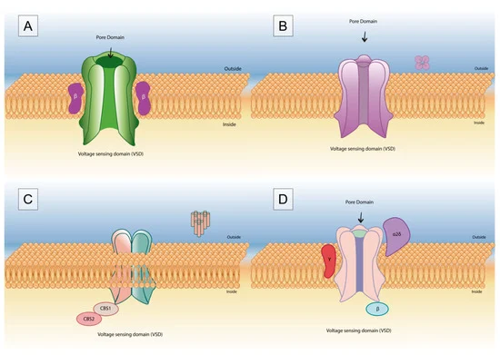 Fig.1 Venom peptides targeting voltage-gated potassium channels.1
Creative Biolabs tailors ion channel screening services to your target to meet your specific venom discovery needs. We provide you with powerful, reliable results, data and timely delivery to accelerate and validate your drug discovery program.
Type of Ion Channels
 Fig.2 Typical ion channels associated with venom peptides.
Our Ion Channel Screening Service for Venom Peptide
Here's a detailed introduction at our ion channel screening services:
-
Traditional Patch-clamp Screening
Conventional patch-clamp technology adsorbs to the cell surface through a special glass tube to form a high-impedance gigaohm seal that accurately records changes in ion channel-mediated current.
- Multiple Rounds of Autocrine Screening
The method is based on an autocrine reporter system that detects the binding of ion channels to toxin peptides. In this system, the venom peptide can be anchored to the cell membrane by the platelet-derived growth factor receptor transmembrane region PDGFR-TM, and the intracellular end of the transmembrane region is fused and expressed with TEV protease. The ion channel is linked to the artificial transcription factor GAL4-VP16 by the TEV protease substrate sequence. When the ion channel binds to the toxin polypeptide, the TEV protease cleaves its substrate to release GAL4-VP16, which activates the expression of the fluorescent protein reporter. When a venom peptide interacts with a potassium channel, cells fluoresce, which is then isolated by FACS (fluorescence-activated cell sorting). Since there will be multiple venom peptide genes in each fluorescent cell, multiple rounds of screening are required.
- High-throughput Venom Peptide Screening Library
The method is built on multiple rounds of autocrine screening, with the introduction of next-generation sequencing (NGS) to establish a venom polypeptide gene library. By synthesizing DNA libraries, and cloning genes into lentivirus. Lentiviruses carry multiple venom peptide genes through viral particles, while different lentiviruses carry some of the same genes. Approximately 1 virion infects a reporter cell. If a peptide introduced by lentivirus reacts positively with a potassium channel within the infected cell, the cell fluoresces red. The red blood cells were then sorted and the venom peptide gene within each cell was amplified and submitted directly to NGS.
High-throughput screening methods enable the discovery of novel venom peptides that block ion channel function. Professional consultation provides guidance before, during and after your venom peptide project, please contact us for your tailored solution.
Reference
- Mendes, Lais Campelo, et al. "Scorpion peptides and ion channels: an insightful review of mechanisms and drug development." Toxins 15.4 (2023): 238. Distributed under Open Access license CC BY 4.0, without modification.
 Download our brochure