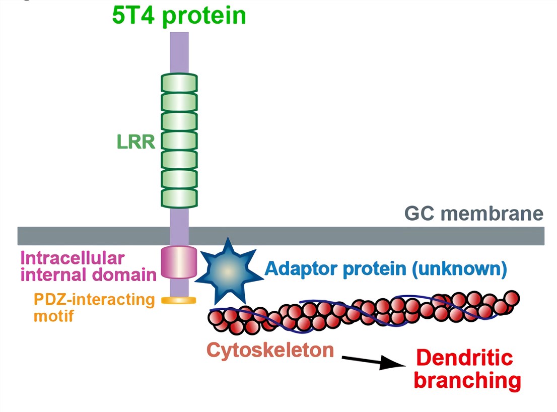
 Fig.1 Schematic representations of 5T4 protein
5T4/WAIF1 (also known as trophoblast glycoprotein, TPBG; 5T4 oncofetal trophoblast glycoprotein and Wnt-activated inhibitory factor 1, WAIF1) is a vertebrate-specific, single-pass transmembrane protein first identified in human placental tissues. Human 5T4/WAIF1 contains 420 amino acid residues and is a single-pass transmembrane protein. 5T4 molecules are 72 kD, heavily N-glycosylated proteins. The extracellular (EC) domain of 5T4 contains seven leucine-rich repeats (LRR) (24 amino acids each), which are often associated with protein-protein interactions. The extracellular part of the 5T4 molecule has 3.5 LRRs in two domains separated by a short hydrophilic sequence with each domain having N- and C-terminal LRR flanking regions. A cytoplasmic domain is a PDZ interacting motif and two serine residues, which are likely phosphorylated by protein kinase Cα (PKCα). 5T4 is present at low levels in other tissues, but high levels in the placenta and most common tumors, typically more than 80% of carcinomas of the kidney, breast, colon, prostate and ovary.
Studies indicate 5T4 and markers of EMT are expressed in cells near the top of the tumor hierarchy implying that 5T4 is possibly associated with EMT. 5T4 was shown to be a marker of the early differentiation of the embryonic stem (ES) cell, and this process involves an E- to N-cadherin switch, upregulation of E-cadherin repressor molecules, gelatinase activity (matrix metalloproteinase (MMP)-2 and -9) and increased cellular motility, all classic EMT features. The cadherins and 5T4 proteins are independently regulated, but studies show that 5T4 and N-cadherin knockout embryonic stem cells show significantly reduced motility during EMT. Moreover, E-cadherin somehow prevents 5T4 cell surface expression and the possible mechanism is through stabilization of cortical actin cytoskeletal organization.
5T4 and C-X-C motif chemokine receptor 4 (CXCR4) molecules are co-localized at the cell surface in differentiating ES cells and mouse embryo fibroblasts which both exhibit C-X-C motif chemokine ligand 12 (CXCL12) specific chemotaxis. 5T4 molecules are involved in the functional expression of CXCR4 at the cell surface in some embryonic and tumor cells. Moreover, 5T4 appears to be stabilizing CXCR4 at the plasma membrane. it is believed that CXCR4 expression facilitates the spread to tissues that highly express CXCL12 to support both the spread and growth of a tumor. CXCL12 is pleiotropic and able to elicit several signal transduction cascades and functions through CXCR4 but also CXCR7. In embryonic cells, it appeared that in the absence of 5T4 expression, CXCR7 is preferentially expressed as the principal receptor for CXCL12 involving transactivation of the epidermal growth factor receptor, which stimulates proliferation or anti-apoptosis rather than chemotaxis. The CXCL12 response outcome is associated with the cell surface 5T4 phenotype. In a tissue/tumor context, it is plausible that surface 5T4 expression at the tumor periphery directs spread towards a local vasculature generated CXCL12 gradient while in the center 5T4 negative tumor cells respond to the chemokine is proliferation or anti-apoptosis.
Wnt protein intracellular signaling is a central component of many aspects of cellular regulation critical to normal development, homeostasis and regeneration, while misregulation can lead to disease, including cancer. It has been shown that 5T4 expression can inhibit the Wnt/β-catenin canonical pathway but at the same time can activate the noncanonical Wnt signaling pathway associated with increased motility. 5T4 interferes with β-catenin signaling in Wnt receiving cells by blocking Wnt-dependent lipoprotein receptor-related protein 6 (LRP6) internalization without affecting low-density LRP6 phosphorylation. At the same time, 5T4 enhances the β-catenin independent Wnt signaling through promoting a noncanonical function of Dickkopf-1 influencing the actin and microtubular skeleton.
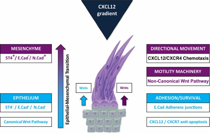 Fig.2 Integrated 5T4 regulation of both chemokine and Wnt pathways. (Stern, 2017)
5T4 is expressed on the cell surface and is present on a wide range of solid tumors (in which expression often correlates with poor prognosis) as a marker of tumor-initiating cells in some cases. Creative Biolabs provides a full set of 5T4 assay portfolio services, such as cell proliferation and growth assay (e.g. BrdU, EDU, CCK8), fluorescent flow cytometry analysis, cytotoxicity assay, in situ hybridization (ISH), immunofluorescence, immunohistochemistry, cell motility assay, migration/adhesion assays, zymogram analysis, ELISA, immunoblotting, immunoprecipitation and so on.
Creative Biolabs is experienced in customizable tumor marker assay services, such as 5T4 assay portfolio service. With hard work over years, we can provide a series of assay portfolio services to assist projects for our customs all over the world. Creative Biolabs offers comprehensive free technical support to ensure your success, please feel free to contact us.
References
For Research Use Only | Not For Clinical Use
 Download our brochure