We provide precise quantitative CEA analysis using highly sensitive and specific methods like ELISA and western blot. Our functional assays, including Cytotoxic T-Lymphocyte (CTL), cell proliferation, and apoptosis assays, investigate CEA's biological impact in vitro.
CEA Assay Portfolio Service
Introduction What We Can Offer Workflow Why Creative Biolabs Customer Reviews FAQs Related Services Contact Us
Creative Biolabs' platform is designed to be highly adaptable, supporting projects from the initial phases of biomarker identification and target validation through to advanced preclinical studies. Creative Biolabs solves key research challenges with a suite of capabilities that includes precise quantitative CEA detection to measure expression levels across various sample types, functional assessment of CEA-related pathways to understand the protein's impact on cellular behavior, and comprehensive support for preclinical studies to evaluate therapeutic efficacy in relevant models.
Functions of CEA
Carcinoembryonic antigen (CEA), also named CD66 or CEACAM5, is a glycophosphatidylinositol -(GPI) linked membrane-anchoring protein. CEA behaves as an intercellular adhesion molecule connecting adjacent epithelial cell membranes, particularly in both embryonic intestine and colonic tumors, resulting in the aggregation of CEA-expressing cells. In addition, CEA plays a significant role in other cellular processes, including the inhibition of differentiation programs, inhibition of anoikis and apoptosis in colon cells, disruption of cell polarization and tissue architecture, and promotion of liver metastasis.
What We Can Offer
Quantitative and Functional Assays
Histological and Cellular Localization
Our immunohistochemistry (IHC) service provides a spatial map of CEA expression in fixed tissue samples. This is crucial for understanding its location, distribution, and relationship to other markers within the tumor microenvironment, which helps guide therapeutic strategies.
In Vivo Validation and Preclinical Support
For advanced research projects, we offer comprehensive in vivo experiments using animal models. These studies bridge in vitro findings to clinical application by assessing the efficacy of CEA-targeting therapeutics, as well as pharmacokinetic/dynamic parameters and safety profiles.
Discover how we can help - Request a consultation.
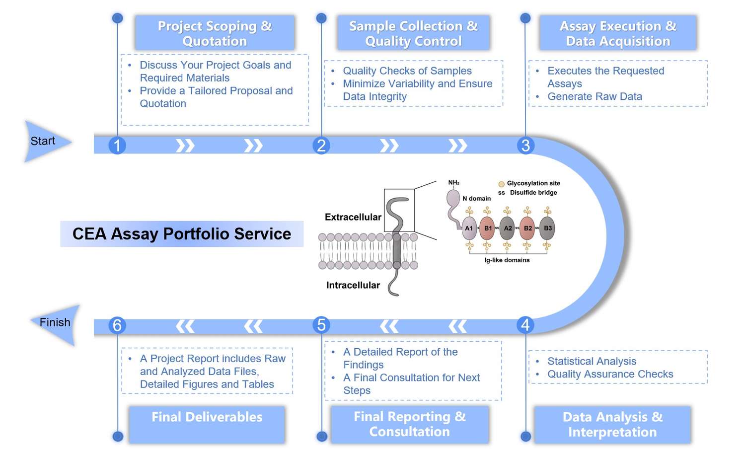
Workflow of CEA Assay Portfolio Service
Highlights
Unrivaled Scientific Expertise
Our team of scientists possesses deep, specialized knowledge of CEA biology, including its roles in cell adhesion, metastasis, and signaling pathways, ensuring our assays address the most critical questions in cancer research.
Comprehensive Service Portfolio
Our full spectrum of CEA assay services creates a seamless research pathway from initial discovery to preclinical validation. This unified model eliminates the challenges of using multiple vendors and ensures data consistency.

Customized Solutions for Every Project
We offer flexible, custom solutions tailored to your unique scientific questions, sample types, and therapeutic molecules. This collaborative process ensures a personalized solution that fits your requirements and accelerates your progress.
Dedicated Partnership and Support
We act as an extension of your team, providing open communication and dedicated support. Beyond data delivery, we offer expert interpretation and strategic guidance, helping you navigate challenges and make informed decisions.
Experience the Creative Biolabs advantage - Get a quote today.
Customer Reviews
-
Seamless Assay Development
Creative Biolabs' expert team guided us through the options and helped us select the most appropriate CTL assay, providing crucial data for our immunotherapy project. - J.S***h, R&D Manager.
-
Exceptional Value and Support
Creative Biolabs' service delivered exceptional value with a lower overall cost, provided superior technical support and consultation throughout the entire project. This enabled us to overcome a key hurdle in our research. - E. L***n, Senior Researcher.
FAQs
What types of CEA assays do you offer?
We offer a comprehensive portfolio of qualitative and quantitative CEA assays. These include core immunoassays like ELISA and western blot for quantification, functional assays such as CTL, cell proliferation, and apoptosis assays to study CEA's biological impact, and in vivo studies for preclinical validation of therapeutics.
Can your services be customized for our specific cell line or animal model?
Yes. We offer custom solutions tailored to each project's unique requirements. Our team works with you to adapt our services to specific cell lines, therapeutics, or animal models, ensuring the data is relevant and actionable.
What is the minimum sample volume required for your services?
The required sample volume depends on several factors, including the specific assay chosen, its intrinsic sensitivity, the expected concentration of CEA in your sample, and the number of technical replicates needed for robust statistical analysis.
Related Services
Antibody and Protein Production
To ensure the highest quality reagents for your assays, we offer a comprehensive recombinant protein production service. This includes the expression and purification of high-quality, full-length CEA protein or specific domains.
Learn More →Immuno-oncology Flow Cytometry Assay Service
This service provides in-depth cellular analysis to characterize cell populations, determine protein expression levels on individual cells, and assess cell viability and function.
Learn More →How to Contact Us
Ready to advance your precision oncology initiatives? Creative Biolabs is dedicated to providing expert-driven solutions that accelerate your scientific discoveries. Our CEA assay portfolio service is designed to be your trusted partner in immuno-oncology research. For detailed information or to discuss your specific project needs, please do not hesitate to contact our team. We look forward to partnering with you on your next breakthrough. Contact us for more information and to discuss your project.
 Download our brochure
Loading case studies...
Online Inquiry