Indoleamine-2,3-dioxygenase 1 enzyme (IDO1) is recognized as one of the most potent therapeutic opportunities to inhibit tumor growth. Multiple studies have shown that IDO1 is highly expressed in several types of human cancer. With advanced and high-end technologies, rich experienced scientists, Creative Biolabs is an excellent service provider in the field of tumor marker assay. After long years ahead to fully comprehend tumor markers, we launch our IDO1 assay portfolio service which can be useful for targeted cancer therapy and diagnosis.
IDO1, a 45 kDa cytosolic haem-containing enzyme, is located on chromosome 8 comprising 10 exonic regions and the promoter region contains several transcription factor-binding sites that have been identified by ChIP-sequencing. IDO1 is distributed in various organs, such as the lung, spleen, liver, kidney and brain, and expressed in many cells, including astrocytes, macrophages and dendritic cells. In addition, Tumor cells can also express widely and variably IDO1, such as the tumor microenvironment's innate immune cells and tumor-draining lymph nodes.
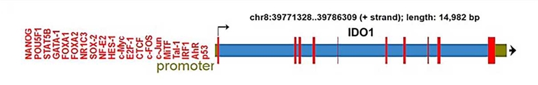 Fig.1 The structure of the IDO1 gene. (Hornyák, 2018)
The expression and function of IDO1 were regulated by multiple upstream pathways, including the JAK-STAT, RAS-PKC, NF-κB, and Kit signaling pathways. The downstream of IDO1 contains three effector pathways that transduce the effects of IDO1 activity, including the activation of GCN2, the inhibition of mTOR and the activation of AhR pathway. The above pathways mediate immunosuppression and neovascularization in the tumor microenvironment.
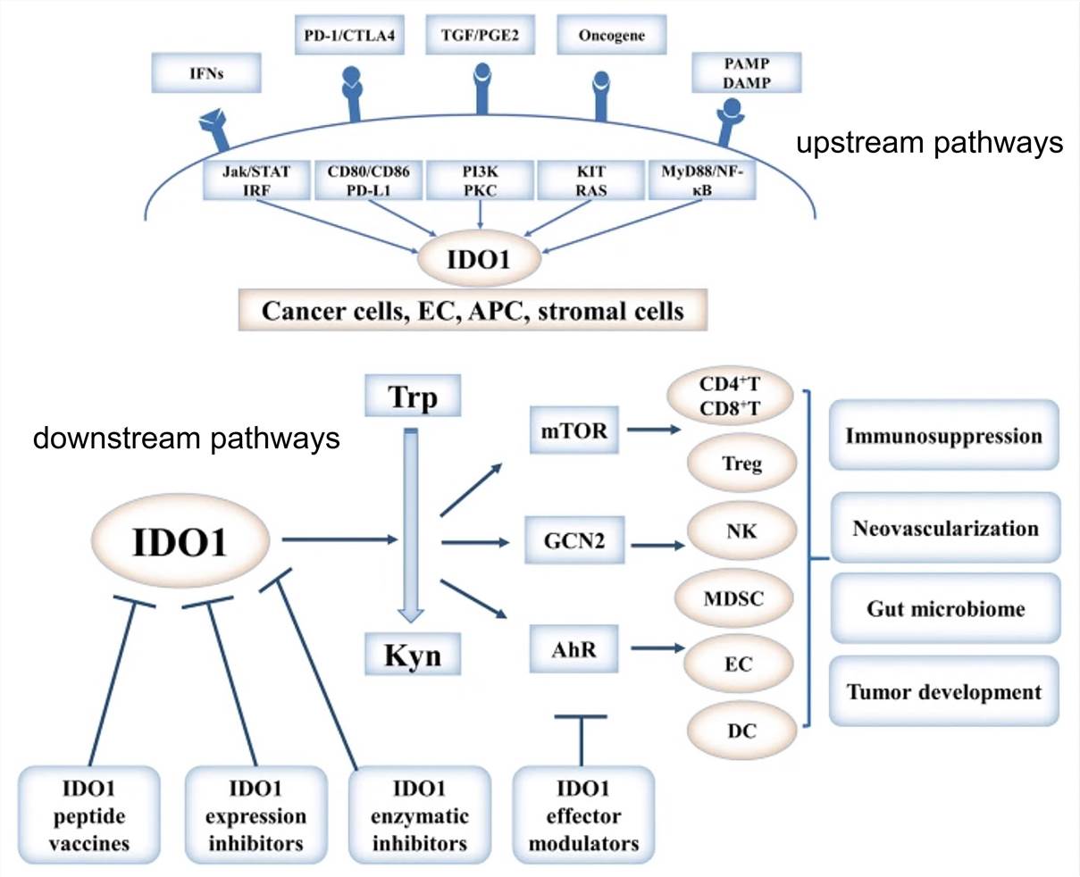 Fig.2 Regulation, function, and targeting of IDO1 in cancer. (Liu, 2018)
IDO1 promotes cancer by forming immune tolerance. It can be attenuated in human tumors resulting from inhibiting by the tumor suppressor Bin1. Additionally, multiple APCs and cancer cells show that overexpression of IDO1 levels can be induced by IL-6, TGF-β, IFN-γ, cytotoxic T-lymphocyte-associated protein 4 and programmed cell death protein 1. IDO1 can enhance the proliferation, invasion, and metastatic abilities of cancer cells to represent a high degree of malignancy in three ways.
If you are interested in our service, please contact us or directly send us.
For Research Use Only | Not For Clinical Use
 Download our brochure