Our assays offer a comprehensive characterization of immune cell subsets within peripheral blood, lymphoid organs, and tumor tissues. We identify and quantify diverse cell populations, which is fundamental for understanding immune modulation and identifying potential biomarkers.
Learn More →Immuno-Oncology Flow Cytometry Assay Services
Creative Biolabs provides cutting-edge flow cytometry assays to enable immunophenotyping and qualification of markers on immune cells. We offer a one-stop solution from consultation about the experiment design, preparation of cells (tissue dissociation, cell isolation, staining, and acquisition), to result analysis. Our analysis instruments have up to 21 colors, which have been widely applied in research settings. This technology can be used in cell counting, cell sorting, identification of various cell populations, and biomarker detection. Moreover, a wide range of cell types is available, including bacteria, yeast, and mammalian cells.
Introduction What We Can Offer Types of Our Services Workflow Why Creative Biolabs Customer Reviews FAQs Related Services Contact Us
Highly Accurate Subpopulation Analysis by Flow Cytometry
Although flow cytometry has excellent performance in detecting rare cell populations and quantifying cellular phenotypes in biological samples, methods of flow cytometric are challenging to develop and validate, especially given the complexity of the samples, the lack of standardized cellular references, and the complexity of measurements involved. However, when designed appropriately, it will provide a wealth of information. Creative Biolabs offers one-stop flow cytometry assay development services, which ensure the whole process can be designed and optimized accordingly.
For a comprehensive discussion of how our capabilities can be leveraged for your specific research, request a consultation.
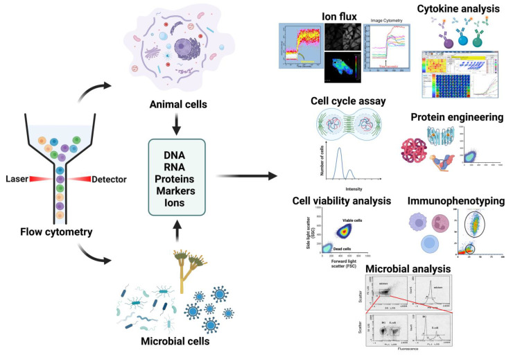 Fig.1 Flow cytometry applications.1
What We Can Offer
Creative Biolabs offers a comprehensive analysis of the immune system.
| Cell profiling and subsetting | Immunophenotyping | Phospho-flow cytometry |
| Intracellular staining | Cytokine measurement | DNA content for cell cycle distribution |
| CAR-T cell characterization | FACS sorting and analysis | Intracellular calcium levels |
| Receptor occupancy (RO) measurement | Spectral flow cytometry | Antibody-dependent cellular phagocytosis assay |
| Phagocytosis assay | Mitochondrial membrane potential | Apoptosis assays |
| Tetramer assay |
Types of Our Services
Immuno-oncology Immunophenotyping Service
Immuno-oncology Receptor Occupancy (RO) Measurement Service
This specialized service quantifies the binding of a therapeutic antibody to its target on immune cells, a critical parameter for determining dosing, scheduling, and efficacy in both preclinical and clinical studies.
Learn More →Immuno-oncology Phospho-flow Cytometry Service
Our phospho-flow service allows for the precise measurement of intracellular signaling pathway activation by assessing the phosphorylation status of key proteins, providing a direct readout of a drug's mechanism of action at the single-cell level.
Learn More →Flow Cytometry Assay Development Services at Creative Biolabs
Highlights
High-Dimensional Insights
Our service provides incredibly rich data sets by leveraging high-parameter multicolor flow cytometry. This technology allows for the simultaneous analysis of numerous markers, bringing a new level of resolution to biological events at the single-cell level, which is both accurate and effective.
Longitudinal Immune Monitoring
Our assays are specifically designed for longitudinal studies and can be used for comprehensive immune monitoring in both preclinical and clinical trials. This allows you to track changes in immune cell populations over time, providing crucial data for understanding therapeutic responses.

Functional Efficacy Analysis
Beyond simple cell identification, our services provide meaningful insights into cellular function. We can analyze key immune cell behaviors, such as natural killer (NK) cell cytotoxicity, to provide direct evidence of your compound's functional efficacy in a highly controlled environment.
Expertise and Precision
Creative Biolabs' over two decades of experience in the field ensure that your project is handled with unparalleled expertise and scientific precision. We develop and validate custom panels and offer advanced data analysis, guaranteeing a reliable and high-quality outcome for your research.
Discover how a partnership with us can streamline your research - Get a quote today.
Customer Reviews
-
Accelerated Antibody Development
Using Creative Biolabs' receptor occupancy service in our antibody development has significantly improved our ability to confirm target engagement in humanized models, saving us time and resources in the early stages. - Aa Ks -
Overcoming Data Complexity
We were struggling with the complexity of our multi-parameter data. Creative Biolabs' data analysis, particularly their use of UMAP to identify novel cell subsets, provided a depth of insight we couldn't achieve internally. - Ml Ro.
FAQs
What is the most important consideration for my samples when using your service?
Proper sample preparation is critical. We recommend a consultation to discuss your specific sample type (e.g., fresh vs. frozen, tissue vs. blood) and handling procedures to ensure the highest data quality.
How do you handle the massive amount of data generated by high-parameter panels?
We use a combination of expert manual gating and advanced computational algorithms to manage and interpret the data. This approach moves beyond subjective analysis and ensures objective, reproducible, and insightful results that reveal the true complexity of your samples.
Related Services
Immuno-oncology Immunophenotyping Service
A holistic service that combines immunophenotyping with other analyses to provide a comprehensive approach to developing cancer immunotherapies.
Learn More →Antibody and Protein Pharmacology
This includes in vivo efficacy, PK/PD, and safety studies for a wide range of therapeutic candidates, including antibodies and proteins.
Learn More →How to Contact Us
Creative Biolabs provides the precision and depth required to navigate the complexities of the tumor microenvironment and make confident, data-driven decisions. Our commitment to quality, expertise in in vivo models, and advanced data analysis make us your ideal partner. Contact us now.
Reference
- Robinson, J Paul et al. "Flow Cytometry: The Next Revolution." Cells vol. 12,14 1875. 17 Jul. 2023. Distributed under an Open Access license CC BY 4.0, without modification. https://doi.org/10.3390/cells12141875
 Download our brochure
Loading case studies...
Online Inquiry