Our advanced discovery platforms leverage cutting-edge bioinformatics and high-throughput screening technologies to help you identify and validate other novel targets for your oncology program.
Learn More →PSMA Assay Portfolio Service
Introduction What We Can Offer Workflow Why Creative Biolabs Customer Reviews FAQs Related Services Contact Us
Creative Biolabs delivers a complete suite of solutions for characterizing your PSMA-targeted compounds. We provide not just data, but a holistic view of your molecule's efficacy and mechanism of action, from initial binding to cellular activity and tissue expression. Creative Biolabs' goal is to de-risk your project and provide the data confidence needed for regulatory submissions and onward development.
Biology and Function of PSMA
PSMA is a transmembrane glycoprotein overexpressed on prostate cancer cells and the neovasculature of many other solid tumors. Its expression levels are correlated with disease progression, making it a highly attractive target for precision oncology. PSMA can enzymatically hydrolyze glutamated folates, producing glutamate and folates that can enter the cell through proton-coupled folate transporter (PCFT), folate receptor (FR), or reduced folate carrier (RFC).
Discover how we can help - Request a consultation.
What We Can Offer
| Functional and Cellular Assays | Protein and Molecular Characterization |
|---|---|
|
|
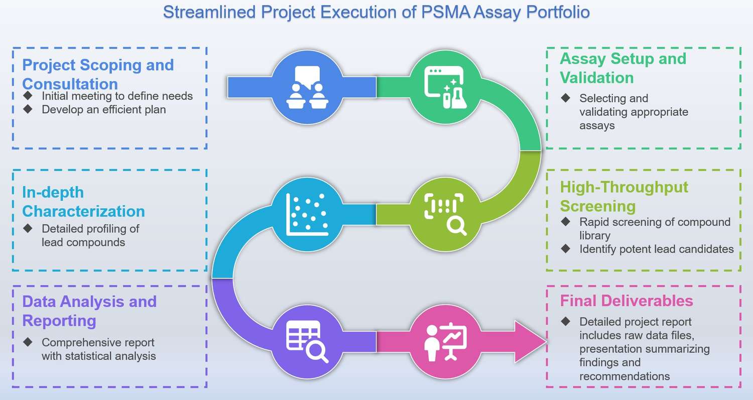
Workflow of PSMA Assay Portfolio
Highlights
Decades of Specialized Expertise
Our deep knowledge base allows us to anticipate and troubleshoot potential challenges before they arise, optimize every step of your experimental design, and provide strategic guidance that helps you navigate the complexities of PSMA-targeted therapeutic development.
Rigorous Quality and Standardization
We guarantee that your results are not only consistent and reproducible but also highly reliable, eliminating the inherent variability and uncertainty that can often plague in-house assay development. This gives you the data confidence required for your most critical milestones.

Holistic Data and Insights
Our comprehensive portfolio allows you to evaluate your molecules from multiple angles, seamlessly connecting biochemical data with functional cellular effects. This integrated approach provides you with a deeper, more robust understanding of your compound's mechanism.
Dedicated Partnership and Support
We are your dedicated partner in discovery. Our team is committed to a collaborative approach, providing expert consultation and interpretation from the initial discussion to the final report, ensuring you have the support you need throughout your entire research program.
Experience the Creative Biolabs advantage - Get a quote today.
Customer Reviews
-
Accelerated Drug Screening
Using Creative Biolabs' PSMA assay portfolio in our research has significantly improved the efficiency. Their HTS platform allowed us to move from a library of thousands of potential candidates to a handful of potent leads in a fraction of the time, shortening our discovery phase. - M*ke.
-
Insight into Cellular Function
The cellular assays offered by Creative Biolabs gave us a much-needed understanding of our compound's mechanism of action in a live cellular context. The apoptosis evaluation via TUNEL assay was particularly insightful and provided critical data. - P*ul.
FAQs
How do you ensure the reproducibility and quality of your data?
All of our assays are developed and validated under strict SOPs, which ensure high levels of consistency and reliability. We implement a tiered QC system that includes plate-to-plate and day-to-day validation, ensuring the robustness of our data. We also provide full transparency with raw data, detailed reports, and a comprehensive analysis of the results, so you have complete confidence in the integrity of our findings.
What is the primary advantage of your portfolio compared to developing my in-house assays?
Our service provides a significant, cost-effective, and time-efficient solution. You gain immediate access to our fully validated, high-throughput platform and our extensive expertise, eliminating the need for a substantial investment in equipment, reagents, and specialized personnel. This allows you to accelerate your research timeline and de-risk your project.
Related Services
Target Discovery and Validation
Cell Line Development Service
This includes the generation of cell lines with inducible or knockdown PSMA expression, as well as reporter cell lines designed for specific functional assays.
Learn More →How to Contact Us
The future of cancer treatment relies on the development of highly effective PSMA-targeted therapies. To achieve this, researchers need a partner they can trust to provide accurate, reliable, and timely data. Creative Biolabs is committed to empowering you with the tools and expertise to accelerate your discoveries, de-risk your projects.
Contact us for more information and to discuss your project.
 Download our brochure
Loading case studies...
Online Inquiry