Viability and Biodistribution Studies
Viability and biodistribution studies provide preliminary data on early tolerance dose, cell survival and CAR/TCR cells kinetic activity of cellualr or gene therapy products. Experimental studies, including elucidating in vivo routes of action, dosing regimens, dose response and utility, and duration of action, provide strong conceptual evidence in selected animal models.
As a leading provider of biotechnology services, Creative Biolabs provides comprehensive pre-clinical viability and biodistribution studies analysis for cell and gene therapy. All tests are performed by experienced technicians using state-of-the-art technology on the most appropriate animal models.
Cell Viability Detection
Cell viability can be measured based on different parameters, such as the redox potential of the cell population, the integrity of the cell membrane, or the activity of cellular enzymes. Each assay provides a different snapshot of cell health and can reflect cell viability individually or together, revealing cytotoxicity or drug efficacy. Creative Biolabs offers a variety of options for cell viability assays:
- Cell lysis or membrane leakage assay: Includes lactate dehydrogenase assay, a stable enzyme commonly found in all cells, which can be easily detected when the cell membrane is no longer intact. Propidium iodide, trypan blue, 7-aminoactinomycin D. and mitochondrial activity or cysteine protease assays (Resazurin and metalaxyl (MTT / XTT) can be used to determine cell death at various stages during apoptosis).
- Functional assay: The measurement of cell function is highly specific to the type of cell being tested. Exercise is a widely used measure of cellular function.
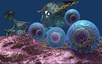
- Genomics and proteomics assays: DNA microarrays and protein chips can be used to measure cellular activation in stress pathways.
Biodistribution Studies
Creative Biolabs offers GLP compliant assessments for the survival of cell or gene therapy products in animal models, tracking their integration in non-target tissues, and the ability to differentiate and migrate at target sites.
For CAR living cells, we can label or stain them in a variety of methods. For example, in cell culture, they can be easily labeled with intracellular dyes such as 5-carboxyfluorescein diacetate succinimidyl ester, 5-bromo-2'-deoxyuridine (BrdU), or green fluorescent protein GFP, followed by in vivo fluorescence imaging.
We Focus On…
- TCR-T and CAR-T/NK/MA cell in vivo proliferation, survival, and persistence
- TCR-T and CAR-T/NK/MA cell migration and distribution in non-target and target sites
- TCR-T and CAR-T/NK/MA cell phenotype analysis
- TCR-T and CAR-T/NK/MA cell response to re-challenge with tumor cells
- Oncolytic virus biodistribution test
For more details, please feel free to contact us for project quotations and more detailed information.
 Download our brochure