B Cell-based Electron Microscopy Assay
Electron Microscopy (EM) for Antibody-antigen Complex Analysis
Electron microscopy (EM) is a high-resolution method that can characterize the structure, conformational states, and subunit compositions of tissues, cells, and complex macromolecules. Electron microscopy is promising in providing crucial structure information as key proof in analyzing molecular function, cell structure changes, and disease. It can even generate 3D structures on atomic resolution level using homogeneous, rigid, biological macromolecules. EM can show three-dimensional conformation and molecular interaction style for dynamic, asymmetric biological molecular complexes involved in critical cellular processes. With advanced instruments and specialized experts in operation, Creative Biolabs offers electron microscopy assay for the determination of the B-cell epitope of cancer antigen, using innovative EM to analyze the binding regions of specific antibodies.
Electron Microscopy Services at Creative Biolabs
Based on the high resolution of EM, Creative Biolabs offers a comprehensive service to directly acquire the structure information on the biological interactions between epitope and monoclonal antibodies, providing essential data on the functional and mechanism study of complex biological processes.
Workflow
Our services cover the following main parts for cancer epitope-antibody analysis. Various methods are available for every step of the whole process. Our experts can provide professional consultation on experiment design and provide strategies that best fit your demand.

Robust Electron Microscopy Platform
The following types of electron microscope are available for applications:
-
Transmission Electron Microscope
-
Scanning Electron Microscope
-
Reflection Electron Microscope
-
Scanning Transmission Electron Microscope
-
Negative Stain Electron Microscope
Our Capabilities
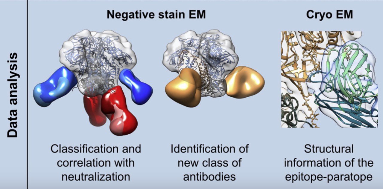
Electron Microscope is an advanced technique used for the confirmation of previous results and data on antigen receptor binding domain (RBD) and determinants of antibodies, providing a 3D structure map of the binding reaction of the complex with nanoscale resolution.
 Fig.1 Functions of EM Technologies on epitope analysis.1
The Function of Electron Microscopy (EM) Assay for B Cell Epitope Analysis
-
Structurally define antibody epitopes and binding regions.
-
Determine the relative location of the binding region to another epitope.
-
Show distinct binding regions of antibody cocktail simultaneously.
-
Confirm the competing antibodies.
-
Describe the mutation map associated with pathogen escape.
Related T and B Cell-based Epitope Analysis Services
In antigen and antibody research, an electron microscope is often combined with other techniques for epitope determination, such as:
Creative Biolabs has developed a reliable T and B-cell assay platform and can also provide assays on monitoring epitope-specific cell responses but not limited to the above. If you are looking for a cancer epitope discovery service, please look at Creative Biolabs. Do not hesitate anymore and contact us right now.
Reference
-
Bianchi, Matteo, et al. "Electron-microscopy-based epitope mapping defines specificities of polyclonal antibodies elicited during HIV-1 BG505 envelope trimer immunization." Immunity 49.2 (2018): 288-300. Distributed under Open Access license CC BY 4.0, without modification.
For Research Use Only | Not For Clinical Use


 Fig.1 Functions of EM Technologies on epitope analysis.1
 Download our brochure

