Fibrosis Biomarker Panel Screening Service
Our Fibrosis Biomarker Panel Screening Service offers a specialized platform for identifying biomarkers associated with fibrosis progression and evaluating potential therapeutic agents. Chronic damage can cause fibrosis, a pathological reaction that disrupts normal tissue architecture and causes an abnormal buildup of extracellular matrix (ECM) components. It is a prevalent characteristic of many chronic diseases, including idiopathic pulmonary fibrosis (IPF), chronic kidney disease (CKD), and liver cirrhosis. It can occur in the liver, lungs, kidneys, and heart, among other organs.
Fibrosis and Biomarkers: An Overview
Fibrosis is characterized by an abnormal wound-healing response, leading to the deposition of collagen and other ECM components in tissues. This process can result from chronic inflammation, oxidative stress, or mechanical injury, and is mediated by various signaling pathways such as TGF-β (transforming growth factor beta), Wnt/β-catenin, and PDGF (platelet-derived growth factor). The progressive deposition of ECM leads to tissue scarring, organ dysfunction, and eventually organ failure.
Fibrosis Biomarker Panel Screening Service at Creative Biolabs
This service provides an in-depth analysis of key biomarkers involved in the fibrotic process, helping researchers identify novel targets, validate biomarkers for clinical use, and evaluate therapeutic candidates in preclinical drug discovery models. Through a combination of high-throughput assays, cellular models, and advanced data analysis, our service accelerates the discovery of effective treatments for fibrosis.
- Biomarker Panel
Our panel covers a broad range of fibrosis-related biomarkers, including but not limited to:
Extracellular matrix (ECM) markers: Collagen types (I, III), fibronectin, elastin.
Fibrosis-related enzymes: MMPs, TIMPs, and lysyl oxidase (LOX).
Inflammatory cytokines and growth factors: TGF-β1, PDGF, IL-6, and TNF-α.
Fibroblast activation markers: α-SMA, vimentin.
Cellular stress markers: HSP70 (heat shock protein), PAI-1 (plasminogen activator inhibitor-1).
- Cell-Based Assays
We use both primary and immortalized cell lines, such as human hepatic stellate cells (HSCs), lung fibroblasts, and kidney fibroblasts, to model the fibrotic process in vitro. These assays can evaluate:
Fibroblast activation: Monitoring markers like α-SMA and collagen deposition in response to profibrotic stimuli.
ECM production: Measuring collagen secretion and ECM remodeling by fibroblasts or other fibrogenic cells.
Inflammatory response: Assessing cytokine secretion or expression of inflammatory markers during fibrosis induction.
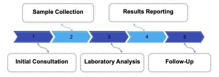
- Service Process
- High-Throughput Screening (HTS)
We offer the ability to screen large compound libraries to identify potential antifibrotic agents. The screening can include small molecules, biologics, or natural product extracts. Compounds are evaluated for their capacity to alter fibrosis-related biomarker expression or activity.
- Mechanism of Action Studies
We can explore the underlying molecular mechanisms of candidate compounds, such as their effects on key fibrotic pathways (e.g., TGF-β/Smad, Wnt/β-catenin, Notch) and their ability to regulate fibroblast-to-myofibroblast differentiation.
- Customizable Panels
Depending on the specific research requirements, we can tailor the biomarker panel to focus on fibrosis in specific organs or tissues (e.g., liver, kidney, lung), or to investigate particular fibrosis subtypes (e.g., idiopathic pulmonary fibrosis, liver cirrhosis).
Unique Features of Our Services
- Comprehensive Coverage: Our panel includes a broad range of fibrosis biomarkers, enabling a comprehensive analysis of both cellular and molecular aspects of the fibrotic process.
- Customizable Approach: We offer flexibility to customize the assays based on your specific research requirements, organ model, or fibrosis subtype.
- High-Throughput Screening Capabilities: The ability to screen large libraries of compounds or biologics ensures that we can identify potential antifibrotic therapies quickly and efficiently.
- Mechanistic Insights: Apart from screening, we offer information on the molecular processes that underlie fibrosis and the possible targets of treatment.
- Expert Data Interpretation: Our team provides detailed reports with data analysis, including biomarker quantification, compound efficacy, and pathway involvement.
Contact Us
If you're ready to explore novel fibrosis biomarkers or evaluate antifibrotic therapies, we invite you to get in touch with us. Our team is ready to discuss your research needs, provide a tailored quote, and offer detailed information on how our services can accelerate your drug discovery process.
 Download our brochure