Metabolism based Fluorescent Imaging Service
Background Service Highlights FAQs Contact Us
Introduction: The Metabolic Imperative in Modern Oncology
For decades, cancer research and therapy have largely relied on anatomical imaging, primarily assessing tumor size as a proxy for disease progression and treatment response. However, a profound understanding of cancer biology reveals that changes in cellular metabolism often precede alterations in tumor morphology. Tumors exhibit a unique and often aberrant metabolic phenotype, characterized by phenomena such as increased glycolysis even in the presence of oxygen (the Warburg effect) and the opportunistic utilization of diverse energy substrates beyond glucose. These metabolic shifts are not merely consequences of uncontrolled proliferation but are fundamental drivers of tumor growth, aggressiveness, and resistance to therapy.
At Creative Biolabs, with over 20 years of specialized expertise in biological imaging solutions, we recognize that to truly conquer cancer, we must look beyond size and delve into the dynamic metabolic landscape of the living tumor. Traditional methods, often invasive and providing only a static snapshot, fall short in capturing these critical, real-time metabolic insights. This challenge has driven our focus on pioneering non-invasive, high-resolution techniques that can visualize and quantify tumor metabolism directly within living organisms. Our commitment at Creative Biolabs is to provide our clients with the cutting-edge tools necessary to unlock these vital biological signals, empowering more precise diagnostics, targeted drug development, and a deeper understanding of therapeutic efficacy.
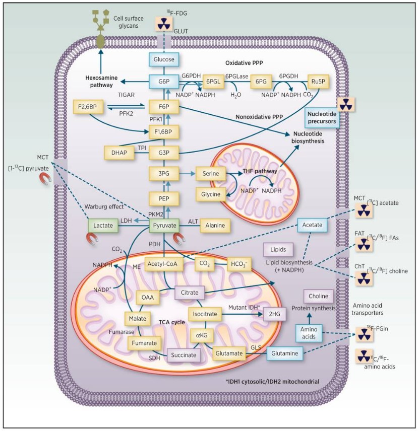 Fig.1 Schematic of clinical imaging for cancer metabolism.1
Our Metabolism-based Fluorescent Imaging for In Vivo Tumor Metabolism Analysis
The intricate metabolic rewiring within a tumor presents both a vulnerability and a diagnostic opportunity. Metabolism-based fluorescent imaging techniques offer a revolutionary approach to non-invasively visualize and analyze these complex metabolic pathways, providing unparalleled insights into tumor behavior and response to interventions. These methods leverage either endogenous cellular fluorophores or specifically designed fluorescent probes to create dynamic "maps" of metabolic activity. Creative Biolabs provides a comprehensive suite of metabolism-based fluorescent imaging services designed to accelerate customers’ oncology research and drug development pipelines.
Fluorescence Lifetime Imaging (FLIM): Endogenous Metabolic Readouts
Fluorescence Lifetime Imaging (FLIM) stands as a powerful label-free technique within the realm of metabolism-based fluorescent imaging. Its principal hinges on measuring the fluorescence decay time of key endogenous fluorophores, primarily Nicotinamide Adenine Dinucleotide (NADH) and its phosphorylated form (NADPH), collectively referred to as NAD(P)H, and Flavin Adenine Dinucleotide (FAD). These molecules are central players in the cellular redox reactions, serving as vital coenzymes in glycolysis, oxidative phosphorylation, and the pentose phosphate pathway (PPP).
Changes in the fluorescence lifetime of NAD(P)H and FAD directly reflect alterations in their binding state to enzymes and the overall redox potential of the cell, thereby acting as intrinsic indicators of the cellular metabolic state. This mechanism allows FLIM to meticulously detect shifts between glycolysis and oxidative phosphorylation, offering a quantitative assessment of metabolic activity at a subcellular resolution. Creative Biolabs utilizes advanced FLIM platforms to reveal subtle variations in metabolic profiles within a tumor, providing a comprehensive understanding of tumor heterogeneity—a critical factor in treatment resistance. Furthermore, monitoring changes in NAD(P)H and FAD fluorescence lifetime serves as an invaluable biomarker for indicating a tumor's early response to treatment, often long before volumetric changes become apparent. This capability extends to both microscopic analysis and whole-animal imaging, furnishing spatial and temporal information about tumor metabolism in a truly in vivo context.
Exogenous Metabolism-based Fluorescent Imaging: Targeted Pathway Visualization
Complementing the label-free approach of FLIM, specific metabolism-based fluorescent imaging techniques utilize exogenous fluorescent dyes engineered to target and label metabolites or integrate into specific metabolic pathways within tumors. This approach provides a powerful means for pathway-specific analysis, enabling researchers to selectively visualize and quantify the activity of distinct metabolic processes.
The mechanism involves fluorescent dyes designed to bind with high specificity to target metabolites or to undergo enzymatic conversion within a metabolic pathway, emitting fluorescence upon binding or transformation. This targeted labeling allows for highly granular analysis of specific metabolic activities, such as glucose uptake, lactate production, or amino acid metabolism. At Creative Biolabs, we leverage these advanced probes to enable real-time monitoring of metabolic changes within tumors, providing dynamic insights into how these pathways adapt under various conditions, including drug exposure. The ability to use different fluorescent dyes to visualize different metabolic pathways is particularly crucial for early-stage drug development, allowing for the precise study of the metabolic effects of novel therapeutic agents on tumors and the identification of potential metabolic vulnerabilities.
Optical Metabolic Imaging (OMI): Rapid Dynamic Assessment
Optical Metabolic Imaging (OMI) encompasses a broader array of optical methods designed to detect dynamic changes in cellular metabolism, including shifts in fundamental processes like glycolysis and oxidative phosphorylation. OMI techniques are founded on the principle that metabolic activity induces detectable changes in fluorescence signals, either from endogenous fluorophores (like NAD(P)H and FAD, as in FLIM) or from exogenously introduced fluorescent probes. The significant advantages of OMI, which Creative Biolabs integrates into its comprehensive service offerings, include its cost-effectiveness, speed, and capacity for directly measuring immediate metabolic alterations. This makes OMI an exceptional tool for high-throughput screening and rapid assessment of metabolic responses in preclinical models.
Our Advantages
Choosing Creative Biolabs for your metabolism-based fluorescent imaging needs means partnering with a leader in the field, backed by decades of collective experience and an unwavering commitment to scientific excellence.
-
Unparalleled Expertise
-
Cutting-Edge Technology & Infrastructure
-
Deep Biological Insights
-
Tailored Solutions for Complex Challenges
FAQs
Q1: What types of tumors or cancer models can be studied using Creative Biolabs' metabolism-based fluorescent imaging services?
A1: Our techniques are highly versatile and applicable to a wide range of solid and liquid tumor models, including patient-derived xenografts (PDX), cell line-derived xenografts (CDX), and genetically engineered mouse models. The non-invasive nature allows for longitudinal studies across various cancer types.
Q2: What specific metabolic pathways can be investigated through these techniques?
A2: We can investigate key metabolic pathways including, but not limited to, glycolysis, oxidative phosphorylation, the pentose phosphate pathway (PPP), and the tetrahydrofolate (THF) pathway. Our use of both endogenous fluorophores (NAD(P)H, FAD) and targeted exogenous probes allows for broad or highly specific pathway analysis.
Q3: How does Creative Biolabs' metabolism-based fluorescent imaging compare to other metabolic imaging modalities like PET?
A3: While PET offers valuable whole-body metabolic information, our fluorescent imaging techniques often provide superior spatial and temporal resolution at the cellular and subcellular level, especially for preclinical models. FLIM, in particular, offers label-free endogenous readouts, providing direct insights into cellular redox states without the need for radioactive tracers. Our services are complementary to PET, offering deeper, more granular metabolic insights within specific regions of interest.
Q4: Is metabolism-based fluorescent imaging suitable for drug screening or preclinical therapeutic efficacy studies?
A4: Absolutely. This is one of the primary applications of our services. By providing early, quantitative biomarkers of metabolic response, our techniques are crucial for assessing drug efficacy, understanding mechanisms of action, identifying drug resistance, and guiding lead optimization in preclinical drug development pipelines.
Contact Us
To learn more about how Creative Biolabs' advanced Metabolism-based Fluorescent Imaging services can accelerate your oncology research and drug development, please contact us today. Our team of experts is ready to discuss your specific project needs and design a tailored solution.
Reference
-
Timm, Kerstin N et al. "Imaging Tumor Metabolism to Assess Disease Progression and Treatment Response." Clinical cancer research : an official journal of the American Association for Cancer Research vol. 22,21 (2016): 5196-5203. DOI: 10.1158/1078-0432.CCR-16-0159. Distributed under Open Access License CC BY 4.0, without modification.
 Download our brochure