Fluorescence Microscopy Imaging Service for Organelle Metabolic Research
Responses of single cells that are undergoing physiological activation processes or pathological stresses are quite heterogeneous in time and space. These parameters in tissue or cellular suspension are usually measured by conventional physiological and biochemical methods. However, these methods fail to resolve discrete differences in the kinetics and magnitudes of the responses between cells, intracellular, and subcellular dynamical processes, which are critical to understanding how cells work.
Scientists at Creative Biolabshave accumulated rich experience in Tumor Subcellular Metabolic Analysis by Raman imaging, MALDI-TOF imaging, fluorescence lifetime imaging (FLIM), and fluorescence microscopy imaging. Regarding fluorescence microscopy imaging, a wide range of indicators sensitive to specific ions and second messenger probes has been developed. Cameleon probes can be targeted to the nucleus, mitochondrial matrix, endoplasmic reticulum, and plasma membrane, while Pericam probes can be targeted to the nucleus, mitochondria, and plasma membrane. The most common way to analyze mitochondrial morphology and dynamics is fluorescence microscopy of mitochondria-targeted fluorescent proteins.
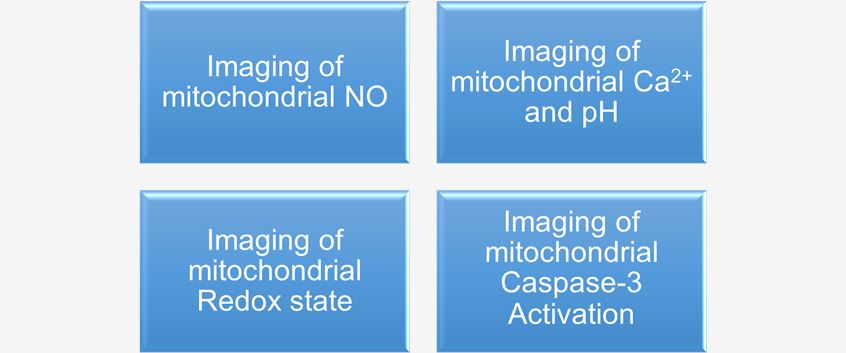
Fluorescence Microscopy Imaging of Mitochondria
Publications Sharing
Publication 1
Technology: Fluorescence microscopy imaging
Journal: Frontiers in Oncology
IF: 4.7
Conclusion: A major contributor to cancer cell survival and proliferation that occurs alongside increased glycolysis activity is metabolism of mitochondria. The role and production of NADH, NADHP, flavins, and various reactive oxygen species (ROS) and how these parameters can be analyzed by fluorescent microscopy are described in this paper. It explains the importance, value, and limitations of label-free autofluorescence imaging of NAD(P)H and FAD. Overall, fluorescence microscopy imaging is an efficient tool to study metabolism in cancer cells.
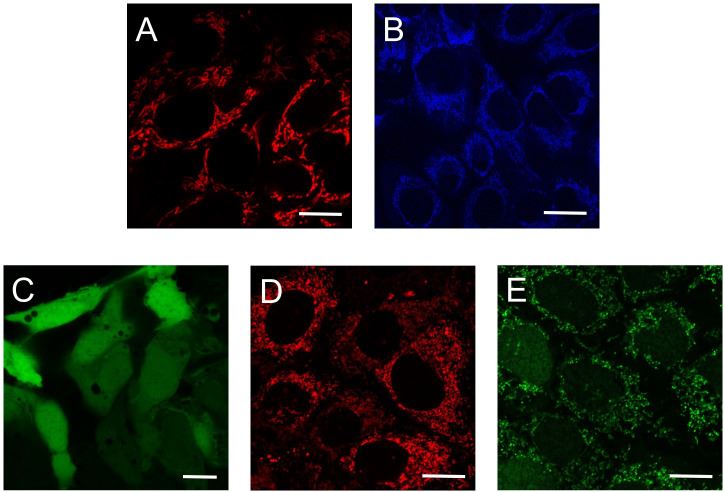 Fig.1 Determination of mitochondrial metabolic activity.1
Except for the Characterization of Tumor Metabolism, we also provide high-quality single-cell tumor metabolic microenvironment analysis, including:
- Single Cell Metabolomic
- Spatial Metabolomics
- Mass Cytometry by Time-of-flight (CyTOF)
- Metabolic Cytometry
- In Situ Dehydrogenase Activity Assay
Our In Vivo Analysis for Tumor Metabolism including:
- In Vivo Isotope Tracing for Tumor Metabolism
- Metabolism-based Fluorescent Imaging
If you have any questions about our fluorescence microscopy imaging services, please contact us.
Reference
- Gooz, Monika, and Eduardo N. Maldonado. "Fluorescence microscopy imaging of mitochondrial metabolism in cancer cells." Frontiers in oncology 13 (2023): 1152553. Distributed under Open Access license CC BY 4.0, without modification.
 Download our brochure